Empowering patients through education—for a life less limited by pain.
Understand your options before you decide
Considering a pain procedure? Start here.
Understand what a treatment can—and cannot—do, how well it may work, its risks, and what recovery could look like. Clear information to help you ask better questions and decide with your clinician.
Education—not a personal treatment recommendation or a promise of pain relief.
Written by a pain physician
Published research cited
No industry funding
Your next step
Start with a treatment guide.
Select a treatment below. Begin with Overview, then use the tabs to explore the evidence, risks, and recovery.
Not sure where to begin? Start with a treatment your clinician has mentioned. The guides help you prepare for that conversation; they do not determine which procedure is right for you.
Your treatment guide
.
Reviewed October 2026 Written by a pain physician, for patients
Educational only — not a diagnosis or treatment recommendation
What Is Spinal Cord Stimulation?
A plain-language starting point. Use the section bar above to go deeper, or swipe left and right on a phone.
Spinal cord stimulation (SCS) is a procedure in which a small device — similar in concept to a pacemaker — delivers mild electrical pulses to your spinal cord. These pulses interfere with pain signals before they reach your brain, reducing how much pain you feel.
It does not fix the underlying cause of your pain. Think of it as changing the volume on your pain signal, not the source. For the right patient, it can meaningfully reduce pain and improve daily function when other treatments have not.
The key distinction
SCS is a reversible treatment. If it doesn't work or you change your mind, the device can be removed. This is one of its most important features compared to surgery that permanently alters anatomy.
Who Is It For?
SCS is generally considered for people who have chronic pain — typically lasting more than 3–6 months — that has not responded adequately to conservative treatments including physical therapy, medications, and injections. The strongest evidence supports SCS for:
Strong evidence
Failed back surgery syndrome (FBSS) · Complex regional pain syndrome (CRPS) · Neuropathic leg pain · Painful diabetic neuropathy
Moderate/evolving evidence
Axial low back pain (without leg component) · Chronic neck pain with cervical radiculopathy · Refractory angina · Peripheral vascular disease pain
SCS is generally not appropriate for
Fibromyalgia · Primary headache disorders · Active cancer as the pain source · Untreated significant depression or active substance use disorder · Patients unwilling or unable to operate the device
The Big Picture Before You Proceed
SCS is a significant decision. It involves two procedures (a trial and, if successful, a permanent implant), carries real risks, requires a psychological evaluation, and demands ongoing device management. Most insurance companies require documented failure of conservative care before they will approve it.
What honest success looks like
SCS rarely eliminates pain completely. The realistic goal is meaningful reduction — enough to improve sleep, activity, and quality of life — not a cure. Published responder rates vary widely because the studies involve different diagnoses, devices, comparison groups, and follow-up periods.
There is no defensible single five-year success percentage for every person receiving SCS. Long-term results should be quoted with the diagnosis, device, outcome definition, and number of patients who were actually followed.
A Brief History of SCS
Spinal cord stimulation is more than 50 years old. Understanding where it came from helps explain both its strengths and its limitations.
1965
The Gate Control Theory — Ronald Melzack and Patrick Wall published a landmark paper in Science proposing that the spinal cord acts as a "gate" that can open or close for pain signals. Large nerve fibers, when activated, could "close the gate" and reduce pain transmission from smaller pain fibers. This was the theoretical foundation for electrical pain treatment.
1967
First human implant — Dr. Norman Shealy, a neurosurgeon, performed the first spinal cord stimulation procedure in a human patient with chronic cancer pain at Case Western Reserve University. The device was rudimentary — a simple electrode placed directly on the spinal cord — but the concept worked.
1970s–1980s
Early commercialization — Medtronic introduced the first commercial SCS systems. Devices were implanted in the epidural space (around the spinal cord) rather than directly on it, reducing risk. Programming was limited and devices were bulky, but the fundamentals were established.
1990s–2000s
Rechargeable batteries, multiple leads, better targeting — Devices became smaller and more programmable. Rechargeable batteries extended device life dramatically. Multiple lead configurations allowed physicians to target pain more precisely. MRI compatibility became an increasingly important design goal.
2010
Burst stimulation (Abbott) — Dr. Dirk De Ridder introduced BurstDR stimulation in 2010, which mimics the brain's own natural firing patterns using a passive-recharge 5-pulse waveform at 40 Hz. Unlike traditional tonic SCS, burst typically works without the patient feeling any tingling — a significant quality-of-life improvement for many patients.
2015
High-frequency 10kHz SCS (Nevro) — The SENZA-RCT, the largest randomized controlled trial in SCS history at that time, demonstrated that 10,000 Hz stimulation outperformed traditional SCS for both back and leg pain, and it required no paresthesia (tingling). This was a major inflection point in the field.
2016
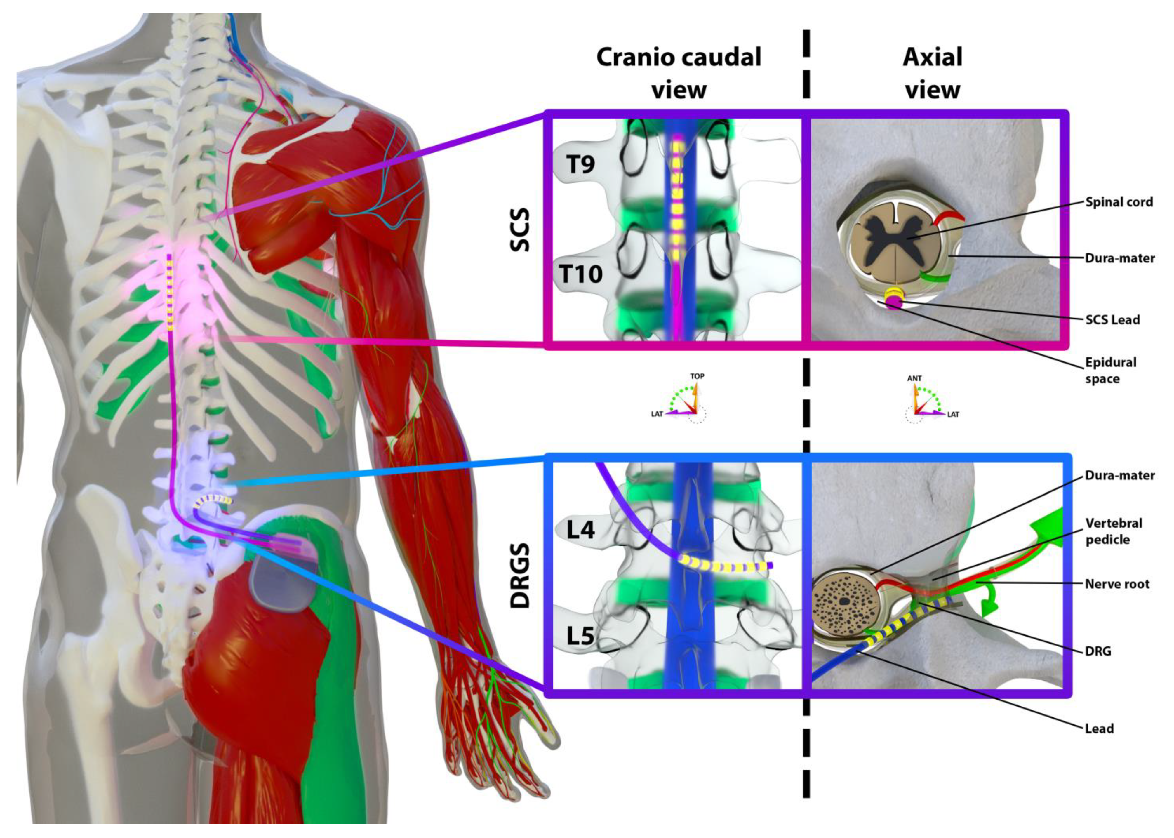
Dorsal Root Ganglion stimulation FDA approval — Abbott received FDA approval for a system that targets the dorsal root ganglion — a cluster of sensory nerve cells that serves as a gateway for specific body regions. This allowed more precise targeting for focal pain conditions like foot pain, groin pain, and CRPS of a limb.
2019–2021
Closed-loop stimulation — Saluda Medical developed Evoke to measure the spinal cord's electrical response and adjust stimulation. The EVOKE trial compared closed-loop with open-loop stimulation using the same platform.
2021
Painful diabetic neuropathy indication — The SENZA-PDN trial demonstrated HF10 SCS was superior to conventional medical management for painful diabetic peripheral neuropathy, opening a major new patient population for the therapy.
Today
App-controlled, AI-optimized, miniaturized — Current systems include smartphone programming, AI-assisted optimization, smaller implants, external battery options, and increasingly personalized therapy. The core concept from 1967 remains, but the technology is unrecognizable.
How Spinal Cord Stimulation Works
The simple explanation has been the same for 50 years. The full explanation is considerably more complex — and that complexity matters for understanding why different waveforms work differently.
The Simple Version: Closing the Gate
Your spinal cord carries pain signals from your body up to your brain. Spinal cord stimulation places electrical pulses near the spinal cord that interrupt or modify this signal before it reaches the brain. Less signal reaching the brain means less pain perceived.
The analogy
Think of a busy freeway carrying pain signals to your brain. SCS doesn't close the freeway — it adds a toll booth that slows and reduces traffic. Fewer signals get through, and the ones that do arrive weakened.
Gate Control Theory (The Original Explanation)
Melzack and Wall's 1965 theory proposed that the spinal cord has a "gate" in an area called the dorsal horn. Large nerve fibers — the kind that carry touch and vibration sensations — can activate this gate and reduce the transmission of signals from smaller pain fibers. Traditional SCS (which produces a tingling sensation) works largely through this mechanism: the tingling you feel is your large fibers being activated, which closes the gate on pain signals.
Why Newer Waveforms Work Differently
Here is where it gets important: high-frequency SCS (10,000 Hz) and some other modern waveforms produce pain relief without any tingling sensation. This means gate control theory cannot fully explain them — the large fibers aren't being activated in the same way. Research suggests these waveforms work through different pathways:
Supraspinal mechanisms
High-frequency stimulation appears to alter pain processing in the brain itself — in areas like the anterior cingulate cortex and thalamus that handle the emotional and sensory aspects of pain. This may explain why HF10 can work even in patients whose pain has a significant central sensitization component.
Neurotransmitter changes
SCS increases the release of GABA (an inhibitory neurotransmitter) and serotonin in the dorsal horn, which dampens pain signaling. It also reduces excitatory neurotransmitters — glutamate and aspartate — that normally amplify pain. The net result is suppression of wide dynamic range (WDR) interneurons: the dorsal horn cells that become hyperactive in chronic neuropathic pain states and drive central sensitization. Calming WDR cells is one of the most important mechanisms by which SCS reduces neuropathic pain at its source. (Burton AW, in Raj's Practical Management of Pain, 4th ed.; Linderoth et al.)
Descending inhibitory modulation
The brain has its own pain-reduction system — pathways that descend from the brain down to the spinal cord and can reduce pain signaling from below. Burst stimulation in particular appears to activate these pathways, essentially triggering the body's own pain management system.
Anti-neuroinflammatory effects
Emerging evidence suggests SCS may reduce inflammation in the nervous system itself — reducing the activation of glial cells (the support cells of the nervous system) that contribute to chronic pain sensitization. This is an active area of research.
What this means for you
The fact that different waveforms work through different mechanisms means there is no single "best" SCS for everyone. The right choice depends on your specific pain pattern, the suspected mechanism driving your pain, and sometimes trial and error. A physician who offers only one waveform type may not be matching the therapy to your biology.
What SCS Does NOT Do
SCS does not repair damaged nerves, fix a herniated disc, reverse arthritis, or treat the underlying structural cause of pain. If you stop stimulation, your baseline pain will return — the device must remain active for ongoing benefit. This is a chronic therapy, not a cure. That is not a failure of the technology; it is simply what it is, and understanding it prevents disappointment.
Understanding Stimulation Parameters — What Your Doctor Is Adjusting
When your physician programs your device, they are adjusting several settings called stimulation parameters. Knowing what these mean helps you give better feedback at programming visits and understand why the same device can feel very different from one setting to another.
Frequency
Hz (Hertz)
Pulses per second
What it is: How rapidly the electrical pulses arrive — measured in Hertz (pulses per second).
The analogy: Think of frequency like the tempo of a drumbeat. Tonic SCS plays a slow, steady beat (40–120 times per second). High-frequency SCS plays it so fast — 10,000 times per second — that it becomes a hum rather than individual beats.
In range: Tonic: 40–120 Hz · HF10: 10,000 Hz · Burst: 500 Hz intraburst · Nalu PSP: up to 1,500 Hz
Amplitude
mA or Volts
Strength of each pulse
What it is: How strong each pulse is — similar to the volume on a speaker. Older voltage-controlled devices measure this in Volts (0–10 V); modern current-controlled devices measure it in milliamperes (mA). Your doctor will know which your device uses.
The analogy: Turn the volume too low and you hear nothing. Turn it too high and it's painful. The "therapeutic window" is the sweet spot — strong enough to reduce pain, quiet enough to be comfortable. Your doctor finds this zone during programming.
Why it matters to you: When you change position (sitting, standing, lying down), the lead shifts slightly and the amplitude can feel stronger or weaker — this is why position-dependent tingling changes are completely normal.
Pulse Width
μs (Microseconds)
Duration of each pulse
What it is: How long each individual pulse lasts — measured in millionths of a second (microseconds). A typical pulse is 100–500 μs.
The analogy: A narrow pulse is a quick tap on your shoulder. A wider pulse is a sustained press. A wider pulse recruits more nerve fibers and spreads the stimulation over a broader area — which can mean better coverage but also stronger sensation.
Why it matters to you: If your tingling doesn't cover your painful area, your doctor may widen the pulse width to expand the coverage — often before changing anything else.
Therapeutic Window
The Sweet Spot
Between too little and too much
What it is: The range between the perception threshold (where you first feel stimulation) and the discomfort threshold (where it becomes uncomfortable). Effective tonic SCS lives in this window.
Good news for paresthesia-free users: HF10, Burst, and DTM work below your perception threshold — you feel nothing, and there is no "too loud" sensation to manage. This makes programming simpler for many patients.
Waveform Pattern
The Shape of the Signal
Not all pulses are alike
What it is: Beyond frequency and amplitude, how the pulses are shaped and grouped matters. Burst delivers 5 rapid pulses in a cluster, then goes quiet. Tonic delivers individual pulses at a steady rate. Each pattern engages different pain pathways.
The analogy: Same notes, different rhythm — a drum hit at 120 BPM feels different than a jazz syncopation at the same tempo. The pattern changes what your nervous system does with the signal.
Impedance
Resistance to Flow
How hard the signal works to reach your cord
What it is: Impedance is the resistance the electrical signal encounters as it travels from the electrode into your tissue. Think of it like water pressure in a pipe — a clog raises resistance.
Why it matters to you: Your doctor checks impedance at every programming visit. High impedance can mean a lead fracture, a loose connection, or scar tissue around the lead. Normal impedance confirms the hardware is working correctly.
Questions to bring to your programming visit
Ask your physician: "What frequency am I running at?" · "Can we adjust the pulse width to cover my [specific area]?" · "Why did my sensation change when I stood up?" · "Is my impedance normal?" Understanding these four questions puts you in a much better position to partner with your doctor in optimizing your therapy.
Waveforms & Devices
Modern SCS is not one therapy — it is a family of related approaches that differ in how they deliver stimulation, what you feel, and what conditions they target best.
How to use this section
Tap any waveform card to expand details. Your physician will recommend a specific approach based on your pain pattern. Knowing the options helps you ask better questions.
Tonic / Conventional Stimulation
All major manufacturers · Oldest technology
Strong Evidence
Frequency
40–100 Hz
Do you feel it?
Yes — tingling sensation
Works via
Gate control theory
Best for
FBSS, neuropathic leg pain, CRPS
In plain English: Your spinal cord has a pain relay station — a layer of cells called the dorsal horn — where pain signals stop before continuing up to your brain. Tonic SCS floods this relay station with a steady electrical hum at 40–100 pulses per second. This overwhelms the "pain routers" (neurons called wide-dynamic-range, or WDR, cells) so they stop forwarding your pain signals. At the same time, it triggers your spinal cord's own chemistry: GABA (the "calm down" chemical) goes up; glutamate (the "alarm" chemical) goes down. Think of it as a constant signal that jams the pain radio frequency. Per Potere et al. in Minimally Invasive Surgical Procedures for Pain (Oxford, 2024), tonic SCS specifically targets the lateral discriminatory pain pathway — the one that tells your brain the location, quality, and intensity of pain.
Why you feel tingling: The same pulses that jam pain signals also activate the touch and vibration nerve fibers running alongside them. That tingling sensation is actually the therapy working — but it means the tingling must physically overlap your painful area for the device to be effective.
Position limitation: When you change position, the lead tips shift slightly, changing how much signal reaches the dorsal horn — so pain relief can vary moment to moment. This was the biggest driver behind newer waveform development.
Burst Stimulation
Abbott (Proclaim XR, Proclaim Plus, Eterna)
Strong Evidence
Frequency
500 Hz (in bursts)
Do you feel it?
Usually no — paresthesia-free
Works via
Descending inhibition, natural firing patterns
Best for
Back and leg pain, back-dominant pain
In plain English: Your brain naturally fires in "bursts" — rapid clusters of pulses followed by silence — especially in the region that governs how emotionally overwhelming pain feels (the anterior cingulate cortex). BurstDR mimics this exact biological rhythm: 5 rapid spikes delivered in quick succession, 40 times per second, with a quiet rest period between each cluster. This pattern mirrors the dual-firing qualities of the thalamus. Most patients feel absolutely nothing — no tingling at all.
The key difference from tonic: Tonic SCS targets the pathway that tells you where pain is and how intense it is (the lateral discriminatory pathway). Burst targets the pathway that makes pain feel emotionally consuming and hard to ignore. Per Potere et al. (2024), burst activates the medial affective ascending pathway — the circuit that drives your brain's attentional and emotional response to pain. This is why burst can help when pain feels overwhelming even when the physical intensity is similar.
Key evidence: The SUNBURST trial (2018) showed ~68% of patients preferred burst over tonic. It may be especially effective for back-dominant pain, historically the hardest target for SCS.
Practical note: No tingling means no easy way to confirm it's working. That's intentional and normal — trust the process and your pain diary.
High-Frequency 10 kHz (HF10)
Nevro (Senza, Omnia, HFX iQ)
Strong Evidence
Frequency
10,000 Hz
Do you feel it?
No — completely paresthesia-free
Works via
Supraspinal mechanisms
Best for
Back and leg pain, painful diabetic neuropathy
In plain English: Chronic pain neurons can get stuck in a "wind-up" loop — firing faster and faster, amplifying themselves into a self-sustaining alarm. At 10,000 pulses per second, HF10 overloads these cells directly at the dorsal horn. Per Potere et al. (2024), this creates a "reversible depolarization blockade" — the pain-processing cells get so saturated with electrical input that they physically cannot fire anymore. It's like tripping a circuit breaker on the pain alarm. When stimulation stops, the block fully reverses — nothing is damaged or permanently changed.
Why no tingling: Unlike tonic SCS, HF10 bypasses the touch and vibration fibers (Aβ axons) entirely and acts directly on dorsal horn pain cells and inhibitory interneurons. No touch fiber activation means no tingling sensation. The textbook also notes HF10 specifically suppresses the WDR "wind-up" phenomenon — the runaway amplification loop that makes chronic pain feel worse over time.
Key evidence: SENZA-RCT (2015): 74% vs. 53% back pain responders; 81% vs. 55% leg pain responders at 24 months vs. traditional SCS. SENZA-PDN (2021): at the 3-month primary endpoint, 79% of painful diabetic neuropathy patients achieved ≥50% relief without worsening neurologic deficits vs. 5% on medical management alone.
Position independence: Because it doesn't rely on touching the right sensory fibers, it works the same whether you're sitting, standing, or lying down.
Boston Scientific Multi-Waveform Platform
WaveWriter Alpha / Alpha Prime · Evidence must be matched to the exact program
Evidence Varies by Program
Output range
2–1,200 Hz
Do you feel it?
Either — felt or paresthesia-free
Main options
FAST, Contour, Illumina 3D, high-rate and burst programs
Platform distinction
Programs can be used separately or in combination
In plain English: Boston Scientific is less a single-waveform system than a toolbox. WaveWriter Alpha can deliver conventional felt stimulation, several paresthesia-free patterns, burst-pattern stimulation, and simultaneous combination therapy. Its Multiple Independent Current Control system steers current in small increments across 16- or 32-source versions, allowing the programmer to shape where stimulation is delivered.
FAST (Fast-Acting Sub-perception Therapy): a low-frequency, paresthesia-free program designed for rapid wash-in. It uses precise targeting and dosing so the response can sometimes be assessed within minutes rather than after a multi-day wash-in.
Contour: a paresthesia-free field-shaping program designed to spread stimulation broadly across multiple vertebral levels and accommodate differences in anatomy or lead position.
Illumina 3D Standard Rate and Prism Targeting: paresthesia-based programming and current steering. The goal is to place comfortable tingling over the painful area; Prism is a targeting tool, not a separate biologic waveform.
High Rate 3D: a paresthesia-free higher-rate option within the platform's 1,200 Hz maximum. It is not the same as Nevro's 10,000 Hz HF10 therapy.
MicroBurst 3D and Burst 3D: configurable burst-pattern choices. These are Boston Scientific programs and should not be treated as identical to Abbott's specifically studied BurstDR waveform.
Combination Therapy: two approaches can run together, such as paresthesia-based stimulation plus Contour. This gives the clinician another programming option before considering a lead or generator revision.
One generator does not mean one evidence grade
WaveWriter is a platform that can deliver several different programs. FDA availability shows the system can deliver them; it does not mean FAST, Contour, Illumina 3D, high-rate, burst-pattern, and Combination Therapy all have the same trials, diagnoses, comparators, or follow-up.
What each study actually supportsMatch the study to the exact program and diagnosis
Combination TherapyContour + felt stimulation together
Direct randomized
COMBO, 89 randomized patients: the three-month composite responder endpoint was met by 36/41 (88%) with simultaneous therapy and 34/48 (71%) with paresthesia-based therapy alone. This supports the tested combination—not every WaveWriter program. Study
Multimodal WaveWriterProgram selected for the patient
Platform randomized
SOLIS, 147 randomized patients with nonsurgical refractory back pain: WaveWriter SCS plus medical management outperformed medical management alone. Because several paresthesia and subperception options were available, the result supports the multimodal treatment strategy in this diagnosis—not one isolated waveform. Study
Subperception ≤1.2 kHzNot a FAST- or Contour-specific trial
Randomized, indirect
WHISPER, 140 previously implanted patients: crossover testing supported noninferiority of subperception to felt stimulation and showed many patients preferred subperception. It does not prove that every named subperception program is equivalent. Study
Rate and neural dosing1, 4, 7, and 10 kHz
Mechanism / dosing RCT
PROCO, 20 selected responders completing randomization: pain relief was similar across rates when pulse width and amplitude were individually optimized; 1 kHz used less charge. This tests rate and dosing in selected responders—not the effectiveness of every commercial program in unselected patients. Study
FAST / FAST-AutodoseFast-acting subperception
Prospective / observational
A prospective single-arm study activated 55 patients and reported 12-month outcomes; an Autodose analysis retrospectively reviewed 73 patients. These results are encouraging but lack a randomized head-to-head comparison with another modern waveform. FAST study · Autodose study
Illumina 3D / ContourField shaping and targeting
Mixed evidence
Illumina 3D: LUMINA was multicenter and observational with a matched comparison, not randomized. Contour: it is part of the randomized COMBO combination, but COMBO cannot isolate Contour’s standalone effect from the paresthesia-based therapy delivered with it. LUMINA
These settings should not inherit results from Abbott’s BurstDR studies. A shared word such as “burst” does not establish waveform equivalence, and no program-specific trial on this page demonstrates that equivalence.
Bottom line: choosing WaveWriter selects a flexible toolbox; choosing FAST, Contour, Illumina 3D, combination, high-rate, or burst-pattern programming selects a different evidence base. Ask: Which exact program will you start with, what diagnosis was studied, what was the comparison group, and was that program tested alone or as part of a package?
In plain English: Chronic pain isn't just a nerve cell problem — it's also a "support cell" problem. Glial cells (microglia and astrocytes) are the maintenance crew of your nervous system. In chronic pain, they become overactivated and act as pain amplifiers, keeping the alarm ringing even when there's nothing left to warn you about. Per Potere et al. (2024), DTM is the only SCS waveform specifically designed to target and reset these glial cells toward a healthier, less sensitized state — essentially working to undo the "pain memory" that chronic pain burns into the nervous system.
How it works: DTM runs four simultaneous electrical signals — one base signal and three "prime" signals, each at different frequencies and pulse widths — targeting six anatomic spots in the spinal cord at the same time. The textbook calls this "multiplexed" stimulation. Think of a symphony conductor directing four instrument sections simultaneously to address the pain orchestra from multiple angles at once, rather than playing one instrument at full volume and hoping for the best.
Evidence note: Randomized DTM studies compared DTM with conventional SCS, not with HF10. DISTINCT studied passive-recharge BurstDR versus conventional medical management; it was not a DTM trial. The glial-cell rationale comes primarily from animal models, so the proposed mechanism is less certain than the clinical pain results.
Closed-Loop / ECAP-Guided Stimulation
Saluda Medical (Evoke) · Medtronic (Inceptiv)
Strong Evidence
Frequency
Variable — auto-adjusted
Do you feel it?
Variable
Works via
Real-time spinal cord feedback
Best for
Patients with variable pain or activity levels
In plain English: Every other SCS device is "flying blind." It delivers pulses and has no idea how much is actually reaching the spinal cord. This matters because when you move — sit up, roll over, lean forward — the gap between your lead tips and the spinal cord shifts. The same setting that gives perfect relief lying down can overshoot when you stand (causing uncomfortable jolts) or undershoot (losing relief entirely). Per Potere et al. (2024), this is the fundamental problem with all open-loop SCS: it provides stimulation without any ability to sense or adjust to the spinal cord's actual response.
What closed-loop does differently: After each pulse, the spinal cord produces a tiny electrical echo called an ECAP (Evoked Compound Action Potential) — a measurable signal that the device can read. Closed-loop SCS listens to this echo thousands of times per second and adjusts the output automatically to maintain a consistent therapeutic level. Think of it as the difference between a fixed-volume radio and one that automatically adjusts for background noise in every room you walk into.
Key evidence: EVOKE compared closed-loop with open-loop stimulation on the Saluda platform. These results should not be assigned to every closed-loop device. Inceptiv uses a separate sensing system; MRI eligibility follows its exact component labeling.
DRG stimulation targets selected sensory nerve ganglia and has a distinct FDA indication and evidence base. Review its trial results, placement, and safety information separately.
Major Manufacturers
These platforms differ in power source, hardware, programming, FDA pathway, and the populations studied. A device's availability does not establish equal effectiveness across diagnoses. Ask which exact device and program your physician recommends, and why.
More patients worldwide carry a Medtronic system than any other brand. Inceptiv is the thinnest rechargeable generator made — about a quarter of an inch — and the family is full-body 1.5T and 3T MRI conditional.
Abbott
BurstDR / FlexBurst360
Key devices
Proclaim XR recharge-freeProclaim Plus recharge-freeEterna rechargeable
Proclaim XR and Plus use a nonrechargeable implanted battery; Eterna is rechargeable. Abbott's .
Nevro
Longest HF10 track record
Key devices
Senza Omnia currentHFX iQ AI-guided programming
Signature technology
HF10 — 10 kHz stimulation, felt as nothing at all
What sets them apart
The company that proved paresthesia-free stimulation works, with the longest commercial track record at 10 kHz. Omnia can deliver any frequency from 2 to 10,000 Hz in one device.
Boston Scientific
Multiple therapies in one platform
Key devices
WaveWriter Alpha currentWaveWriter Alpha Prime currentSpectra WaveWriter earlier platform
Signature technology
FAST · Contour · Illumina 3D · High Rate 3D · MicroBurst / Burst 3D · Combination Therapy
What sets them apart
One generator can provide both felt and paresthesia-free programs and can run selected therapies together. The 16- and 32-source versions use independent current control for detailed field shaping. WaveWriter Alpha is rechargeable; Alpha Prime is recharge-free. There is no single platform-wide responder rate: COMBO, SOLIS, WHISPER, PROCO, FAST, and LUMINA studied different programs, populations, designs, and comparators.
Nalu (Boston Scientific)
No internal battery
Key devices
Nalu Neurostimulation System
Signature technology
PSP (Pulsed Stimulation Pattern) · Tonic · Micro-generator powered by a wearable patch
What sets them apart
There is no battery inside you, so there is never a battery-replacement surgery. The generator is about the size of a large coin; power comes through the skin from a charger worn like a patch. Nalu Medical was independent until Boston Scientific completed its acquisition in January 2026, so you may see it listed either way.
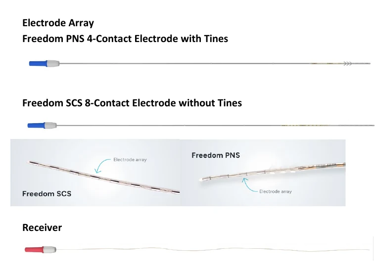
Curonix — Freedom SCS
Externally powered · FDA cleared
What is implanted?
Electrode arrays and receivers. No implanted battery or conventional IPG.
What you wear
A rechargeable transmitter and antenna held in a torso wearable supply energy during stimulation. This makes the wearable part of the daily treatment.
Programming
HF-EMC describes the wireless power link. It does not mean the stimulation is automatically 10 kHz. Ask which stimulation patterns are permitted by your exact system's labeling.
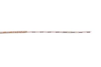
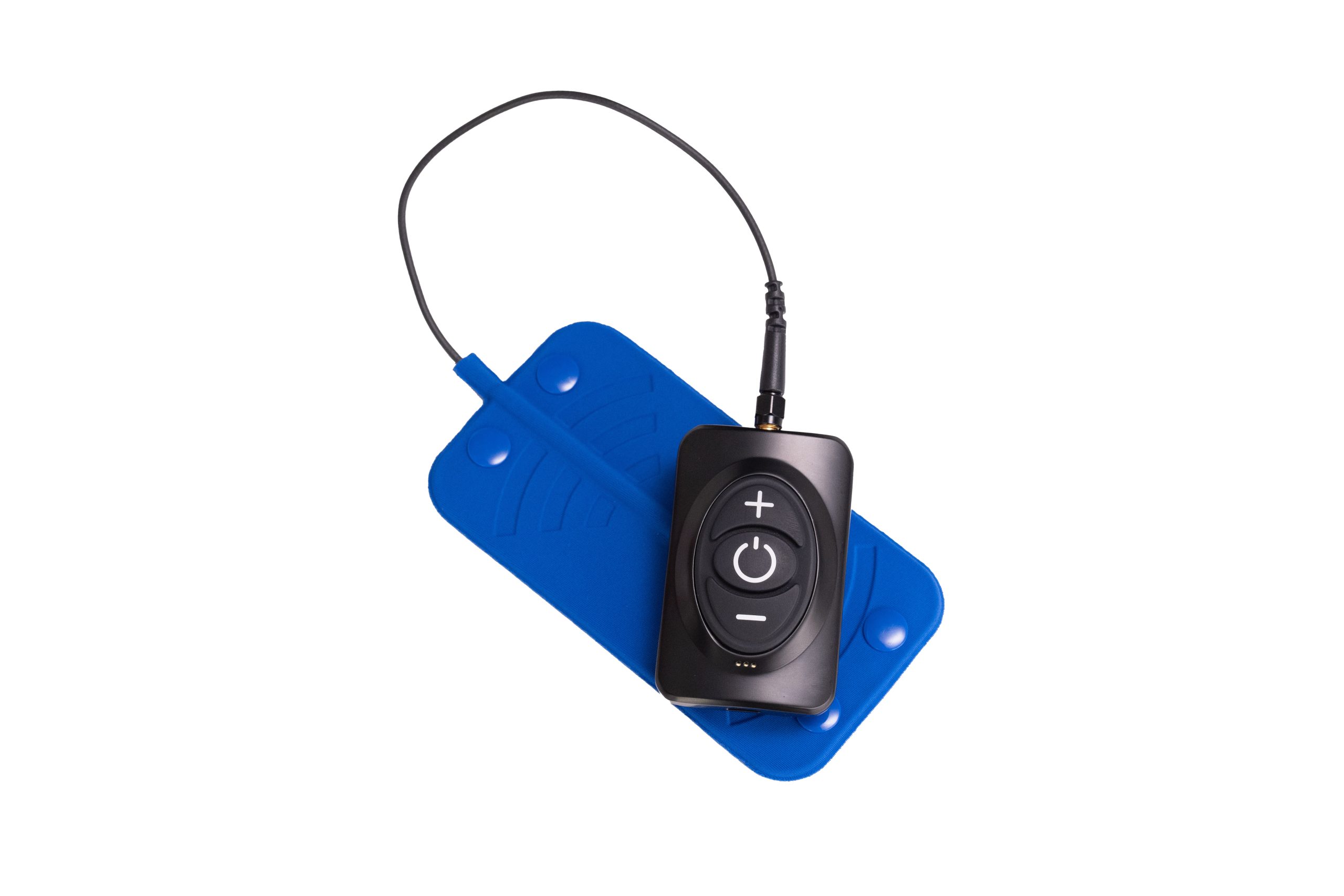
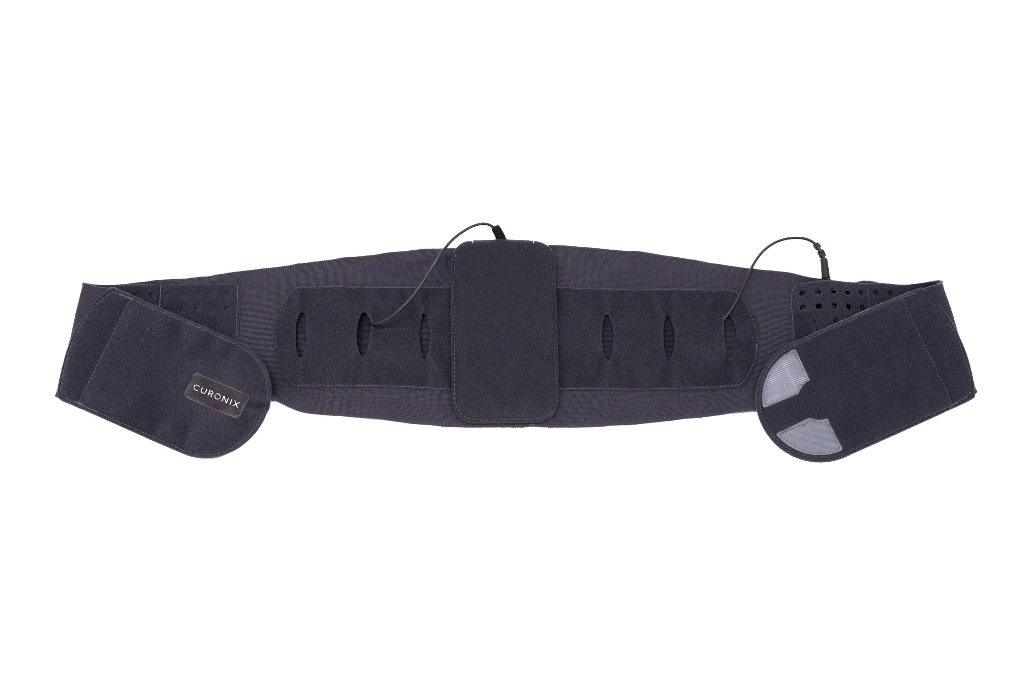
These manufacturer photographs show the three parts to recognize: the implanted electrode array, the external power assembly, and the torso wearable. Images are shown at different magnifications.
Under the skin: electrode array connected to an implanted receiver. FR8A: 8 contacts, 1.35 mm lead diameter, 45 cm lead length.Outside the body: rechargeable transmitter and antenna deliver power and programming through the skin.During treatment: the torso wearable holds the power assembly in position, over a layer of clothing.
Usually no power wearable; rechargeable models need charging sessions
Therapy disc over the micro-IPG
Transmitter + antenna in torso wearable
Practical question
Where will the generator sit, and how often will I charge or replace it?
Can I comfortably wear and align the therapy disc?
Can I comfortably position and wear the power assembly whenever therapy runs?
Curonix evidence: match the study to the claim
SURF (2022) reported at least 50% back-pain reduction in 33/39 completers (85%) at six months. It was a small prospective single-cohort study, excluded trial failures, and had no sham or competing-device arm.
A 2019 randomized study compared two waveforms on the same Freedom hardware; it did not compare Curonix against another manufacturer. Those results do not establish a universal success rate or superiority over other SCS systems. SURF · Waveform RCT.
IPG Size & Implant Location Comparison
The same-scale lineup below compares generators. Curonix is shown separately above because its implanted array/receiver and external power hardware do not form a conventional battery IPG.
The implantable pulse generator (IPG) — the battery and computer — varies significantly in size across manufacturers. Smaller is more comfortable for thin or active patients; larger devices generally hold a bigger battery or support more features.
Every generator at the same scale
Width × height × thickness, in inches. A U.S. quarter sits at the left for reference.
Swipe sideways to see every device →
Ordered from smallest to largest. Notice the trade-off between footprint and thickness: Medtronic’s Inceptiv is the thinnest (about ¼ inch) but is not the smallest footprint; Abbott’s Eterna has the smallest footprint but is thicker. Device illustrations are original, drawn from each manufacturer’s published dimensions — not product photographs. Quarter: U.S. Mint photograph (public domain).
Nalu Medical
0.19 in thick
Smallest available — about the size of a large coin · 1.10 × 0.43 × 0.19 in
Battery-free micro-IPG. No internal battery means no replacement surgery ever. Power comes from an external wearable charger worn on the skin.
Typical location: Flexible placement due to tiny size. External charger wears like a patch.
Abbott — Eterna
0.35 in thick
Compact rechargeable — about the size of a matchbook · 1.89 × 1.52 × 0.35 in
Supports BurstDR, tonic, and sub-perception waveforms. One of the smaller full-featured rechargeable IPGs.
Typical location: Upper buttock or lower abdomen/flank.
Largest — ECAP sensing hardware · 1.89 × 2.68 × 0.47 in
Extra space accommodates sensing hardware for real-time closed-loop ECAP feedback — the most advanced automated dosing currently available.
Typical location: Upper buttock. Most relevant size concern for thin or petite patients.
Where is the IPG placed?
The two most common sites are the upper buttock (posterior superior gluteal area) and the lower abdomen or flank. Your surgeon will choose based on body habitus, dominant hand, and lead routing. The abdomen is sometimes preferred because bending at the waist creates less mechanical stress on the lead-to-IPG connection.
The IPG must be reachable with your dominant hand to use the patient remote. Discuss placement preferences before surgery — it is much easier to choose the right spot in advance than to revise it later.
Lead Differences Between Companies
The leads are thin wires that deliver electrical pulses to your spinal cord. Lead design — how many contacts, what shape, and how they're placed — significantly impacts which areas can be treated and how precisely therapy is delivered.
The two kinds of leads
Drawn at twice the scale of the generators above; the quarter is enlarged to match, so the comparison still holds.
〰️
Percutaneous (Cylindrical)
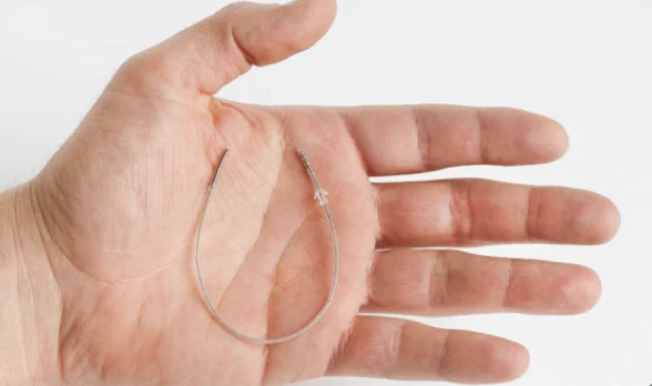
Thin, flexible wire inserted through a needle — no incision needed. Contacts ring the circumference of the wire.
✓Placed during office trial — no OR needed
✓Can be repositioned without surgery
✓Smaller procedure, faster recovery
✗More prone to migration (moving)
✗Less precise dorsal column coverage
▬
Paddle (Surgical)
Flat lead placed via small open surgery. Contacts face only toward the spinal cord — more targeted and efficient stimulation.
✓Highly stable — very low migration rate
✓Better dorsal column coverage
✓Lower energy → longer battery life
✗Requires surgery and anesthesia
✗Cannot be easily repositioned
Lead Contacts & Key Features by Manufacturer
Company
Lead examples
Contacts per lead
Placement / model notes
Abbott
Octrode; Penta; Lamitrode family
Octrode: 8 Penta: 16 Lamitrode: model-dependent
Needle or surgical paddle, depending on model. Penta belongs to Abbott. Labeling
Medtronic
Vectris; Specify 2×8 / 5–6–5
Vectris: 8 These Specify models: 16
Percutaneous or paddle. Exact component combination controls MRI eligibility. See model-limited recall notes below. Manufacturer report
“Illumina” is programming technology. Infinion CX SC-2317-50/-70 has a 2026 Class I recall; do not treat all Infinion variants as identical. Labeling
Nevro
Percutaneous leads; Surpass / Surpass-C paddles
Percutaneous: 8 Surpass: 16
The standard Surpass paddle has two columns. “Tripole” belongs to Abbott; the earlier 24-contact Nevro claim was incorrect. Surpass manual
Saluda
Evoke 12C; CAP24 paddle
12C: 12 CAP24: 24
Stimulate and sense ECAPs. CAP24 expanded the surgical paddle options in 2026. Manufacturer announcement
Nalu
Nalu percutaneous lead models
Model-dependent; check implant card
Leads connect to a battery-free micro-IPG powered by a therapy disc. The 2024 notice below concerned certain IPG kits. FDA record
Curonix
Freedom SCS FR8A array + receiver
8
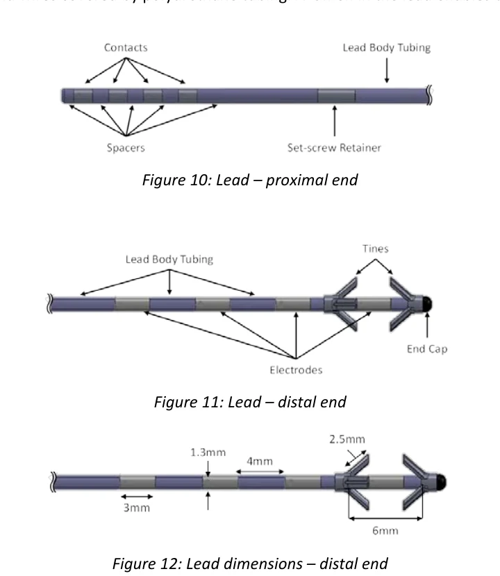
1.35 mm diameter; 45 cm lead; 3 mm contacts with 4 mm spacing. External transmitter supplies power. SCS instructions
SCS Lead Recalls & Device Safety Notices
Reviewed October 20, 2026 against FDA records and manufacturer notices. Match the exact model, lot/PIN, and serial number on your implant card; a family name alone cannot determine whether a notice applies.
Boston Scientific Infinion CX: 2026 Class I recall
SC-2317-50 and SC-2317-70. Stress near the anchor can cause high impedance or lead fracture, loss of therapy, and possible revision. The June 17, 2026 action removes unused stock.
The FDA notice says this action does not affect already implanted devices and recommends no extra monitoring beyond routine clinical follow-up. A recall does not automatically mean an implanted lead should be removed. Discuss changed or lost stimulation with your treating team. FDA patient notice · Recall record.
Other model-specific and historical notices
Device
What was affected?
Scope / source
Medtronic Vectris SureScan MRI
2021 Class II: electrode-spacing labeling. Models 977A260/275/290; PINs 977A20024V/25V/28V.
Open/classified in FDA record. Listed distribution: Europe/Brazil. This is a limited labeling action. FDA
Medtronic Specify 39565 / 39286
2016 labeling correction about trial versus permanent use.
2024 Class II: RF efficiency / IPG manufacturing issue in 13 serial-limited kits.
Open/classified. This concerns certain IPG kits, rather than a lead-fracture recall. FDA
Abbott Proclaim SCS / DRG and Eterna
April 2026 Surgery Mode correction for Proclaim; October 2025 Bluetooth communication advisory for Eterna 32400.
Generator / system handling notices; separate from a lead recall. Abbott advisory index
Other displayed lead examples
No matching lead-specific recall identified in this review for Abbott Octrode/Lamitrode, Curonix Freedom SCS FR8A, Saluda Evoke 12C/CAP24, or Nevro percutaneous/Surpass leads.
This is a search finding, not a guarantee about every model, lot, or future notice. Check the implant card against the FDA recall database.
These are the relevant records identified in this review, not a certification that every other model or lot is recall-free. For a planned implant, ask the team to confirm the exact components against current notices.
What does "more contacts" mean for you?
More contacts give your programmer more flexibility to customize where stimulation is delivered. This matters most if your pain covers multiple areas (both legs and low back), or if initial programming doesn't relieve all your pain. More contacts ≠ better outcome — it mainly increases flexibility for complex cases.
A note on device company marketing
All major SCS manufacturers invest heavily in marketing to physicians and patients. Clinical trial data is often funded by the device manufacturer, which doesn't invalidate the data but does mean independent replication is important. When your physician recommends a specific device, asking "do you have a financial relationship with this company?" is a fair and legitimate question.
Which Waveform Is Right for This Patient?
An evidence-based decision guide — for patients and providers alike. Answer three questions to see which waveform the research most strongly supports, and exactly why the others weren't chosen.
1
2
3
Are you a patient or a healthcare provider?
This adjusts the language so it's most useful for you.
The Spinal Cord Stimulator Trial
A temporary, real-life test of pain relief, function, and whether the therapy fits your daily life—before anything permanent is implanted.
What the trial is really testing
The trial asks, “Is this therapy worth pursuing?” It is not a miniature permanent implant and it does not guarantee the long-term result.
The trial weekTemporary leads; external generator
Usually 5–7 days
This is a short, real-life test—not a promise of the permanent result. The taped dressing and outside battery are awkward, and the insertion site may be sore, but there is no implanted battery pocket to heal.
Purpose
Test pain relief, function, sleep, and whether using the system feels manageable.
At home
Keep the dressing and external equipment dry. Follow the team’s limits on bending, twisting, lifting, and reaching so the temporary leads do not move.
Contact team
Call during the trial if coverage is poor or uncomfortable. A programming change may be part of a fair test.
Removal
The temporary leads are usually pulled in clinic. If the trial helped, permanent implantation is scheduled separately.
During lead placement
Often awake, but comfortable
Numbing medicine plus light intravenous sedation is common. You are drowsy and comfortable but may be asked where you feel stimulation while the physician positions the lead.
Sedation practice varies by center and waveform. Ask your physician exactly what to expect and whether you will need to provide feedback.
How to use the trial as a real testDo ordinary, safe activities within your procedure restrictions. Do not “stress test” the leads with bending, heavy lifting, or strenuous exercise.
BeforeChoose 2–3 goals
Examples: stand through a shower, walk to the mailbox, sleep four uninterrupted hours, or sit through a meal.
Day 1Learn the system
Record baseline pain and procedure soreness separately. Confirm how and when to contact the programmer.
Days 2–6Repeat and record
At the same times each day, note target-pain relief, goal performance, sleep, medicines, and side effects.
Last dayDecide with evidence
Review the diary with the clinician. A memorable good hour should not outweigh the whole week.
What usually counts as a successful trial?
The 2023 evidence-based consensus guideline recommends documenting at least 50% improvement in pain with a validated measure and assessing a patient-specific functional goal. A decision can be individualized when function or medication use improves substantially, but insurer rules differ.
PainCommon benchmark: at least 50% relief in the specific pain being treated—not necessarily every pain.
FunctionCould you do a personally important activity farther, longer, or with less recovery afterward?
FitWas the stimulation tolerable, and could you use the controller and charging system expected for that device?
72–82%reported median trial success
Across randomized and observational reports summarized by the 2023 consensus panel.
61–65%reported 12-month therapy success
These are different evidence pools—not a direct “drop-off” calculation and not an individual prediction.
A successful trial does not guarantee the same long-term result
The temporary and permanent systems differ, early implant soreness can blur the comparison, and benefit can change with healing, lead position, programming, diagnosis, or the pain itself. Judge the permanent system after healing and reasonable programming—not from the first postoperative day. Sources: 2023 consensus guideline and Johns Hopkins patient guide.
The Process — Step by Step
From first appointment to long-term management, here is the full SCS journey at a glance. Use the separate The Trial and Permanent Implant tabs for the practical details of each procedure.
How the device is placed
Three stages, from lying down on the table to the finished implant.
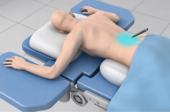
1 You lie face-down
On your stomach on the table, with a cushion under your hips to open up the spaces between the vertebrae. The skin over the entry point is numbed.
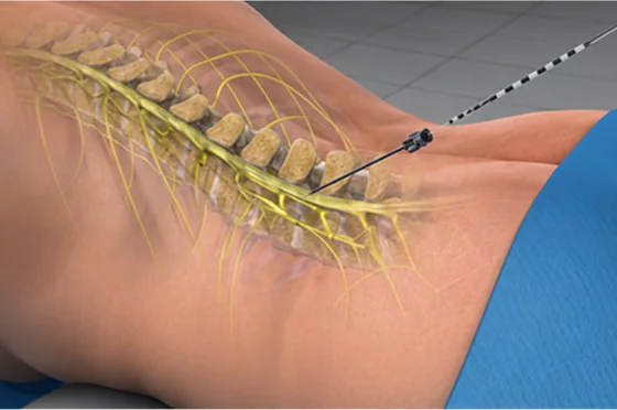
2 The lead goes in through a needle
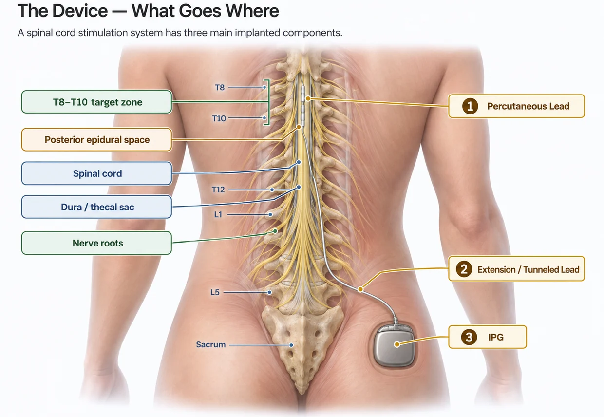
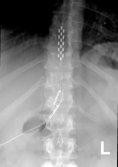
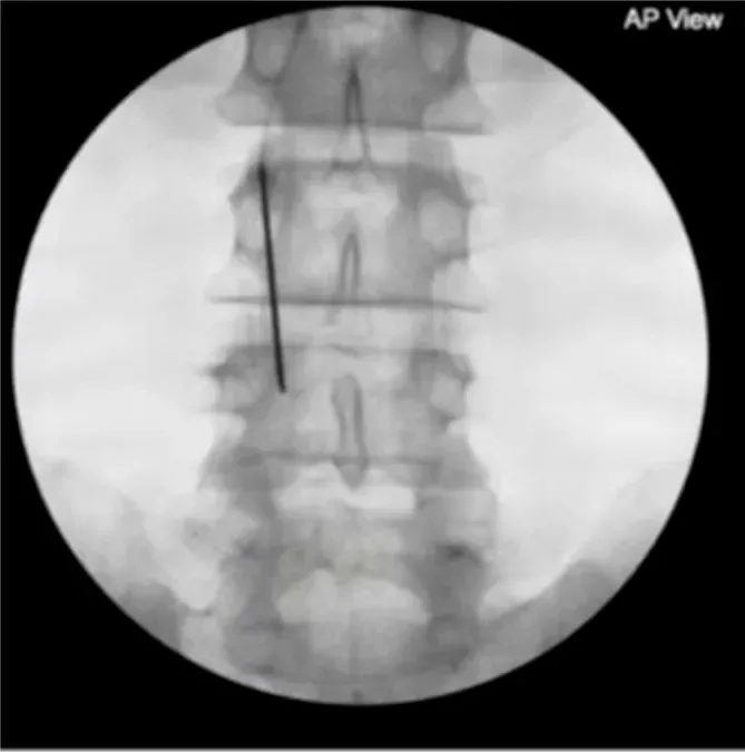
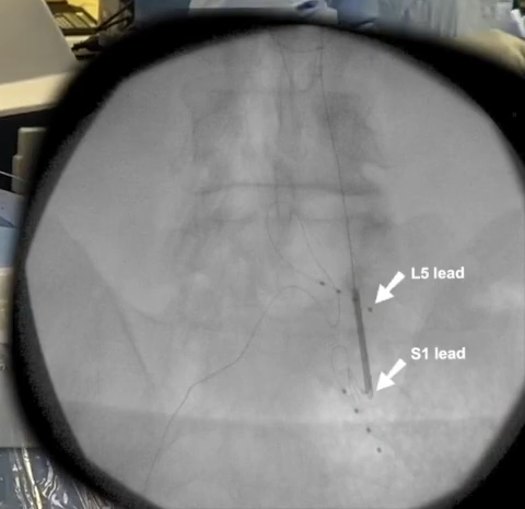
No cut is made. A needle is guided into the epidural space using live X-ray, and the thin lead is threaded up through it to sit behind the spinal cord.
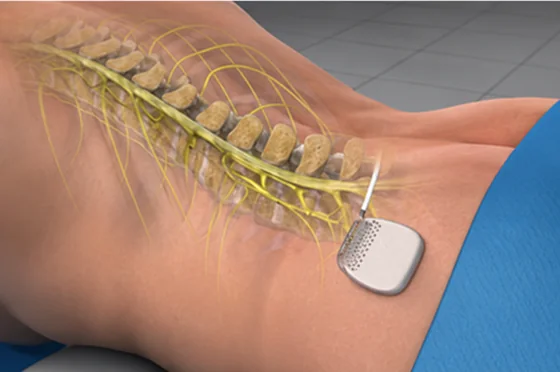
3 The generator goes under the skin
Only at the permanent implant. The leads are tunnelled under the skin to a small pocket that holds the generator. For the trial, the wires simply exit the skin instead.
The needle goes in low, around L2–L3, below where the spinal cord ends — then the lead is threaded upward to sit over the cord at T10–T12. That is why the entry point and the treatment level are different places.
1
Weeks 1–4
Evaluation & Candidacy
Your pain physician reviews your history, imaging, prior treatments, and performs a physical exam. Key questions: Have you completed adequate conservative care? Is your diagnosis appropriate for SCS? Are there any contraindications (pacemaker, active infection, anatomy that prevents lead placement)?
Psychological evaluation
Most physicians and virtually all insurers require a psychological evaluation before SCS. This is not a barrier or a test to pass — it identifies factors that predict better outcomes (active coping, realistic expectations) and factors that predict worse outcomes (untreated depression, opioid dependence, catastrophizing). A good physician sees this as part of your care, not a box to check.
2
Weeks 4–16 (most variable stage)
Insurance Pre-Authorization
Your physician's office submits a prior authorization request to your insurance company. This includes your diagnosis, imaging, documentation of failed conservative care, medication trials, and the psychological evaluation. Timelines range from 2 weeks to 4 months depending on your insurer.
What causes delays
Missing documentation of conservative care failure, psychological evaluation not yet completed, or diagnosis code mismatch. Stay in contact with your physician's office — ask for the prior auth tracking number and check status every 2 weeks.
3
Day 1 — Outpatient procedure
The Trial
The SCS trial is a temporary test. Thin, flexible leads are usually placed through a needle into the epidural space under fluoroscopy (live X-ray guidance), then connected to a small external battery. Most published studies use a 5–7 day trial; the exact duration, anesthesia, and dressing instructions vary. You usually go home the same day.
Your job during the trial
Keep a daily pain diary. Rate your pain morning and evening. Note which activities you can do that you couldn't before. Most physicians use ≥50% pain relief as the threshold for a successful trial. What matters most is whether the relief is meaningful to your daily life.
4
Day 7–10
Trial Removal & Decision
The temporary leads are removed in your physician's office. You and your physician review your trial diary and discuss whether to proceed.
If successful: Proceed to permanent implant — insurance requires documentation of trial success.
If unsuccessful: Leads are removed, nothing permanent has been implanted, and you explore other options. This is an important patient protection — the trial requirement prevents permanent implants in patients who won't benefit.
5
2–6 weeks after successful trial
Permanent Implant
Done in an operating room or ambulatory surgery center with sedation or general anesthesia, depending on the patient, lead type, and center. Permanent leads are placed and the IPG (internal pulse generator—the battery/computer) is implanted under the skin, commonly in the buttock, flank, or lower back. Many patients go home the same day, while a more involved paddle-lead operation may have a different recovery plan.
Percutaneous vs. paddle leads
Percutaneous leads (through a needle, like the trial) are minimally invasive but have higher migration rates. Paddle leads (placed surgically through a small laminotomy) sit more stably but require a larger procedure. Your physician will recommend based on your anatomy and history.
6
First 3–6 months post-implant
Programming & Optimization
The device is programmed at your post-operative visit. Most patients require 2–4 programming sessions over the first few months. Think of it like adjusting a new hearing aid — the initial settings are a starting point, not a final answer. Modern systems increasingly allow remote adjustments via smartphone app.
7
Ongoing — years
Long-Term Management
Rechargeable IPG: Charge daily to weekly using a pad placed over the implant site. Battery life 7–10+ years before replacement surgery.
Non-rechargeable IPG: No charging, but requires surgical battery replacement every 3–7 years.
MRI: Most modern devices are MRI-conditional with specific precautions. Always tell any imaging facility, emergency room, or treating physician that you have an SCS implant before any procedure.
The Permanent Implant
What usually happens on implant day, how recovery differs from the trial, and when the device is adjusted for long-term use.
From procedure day through the first six weeks
The permanent implantLeads and generator under the skin
Healing takes weeks
This is a larger procedure than the trial. With percutaneous leads, there is usually a lead-anchoring incision plus a pocket for the battery. A paddle lead requires a more involved surgical approach.
Procedure
Often outpatient and roughly one to two hours, but anesthesia, lead type, and hospital stay vary by patient and center.
First days
Incision and battery-pocket soreness are expected. That new surgical pain is different from the chronic pain the device is meant to treat.
Weeks 1–2
Wound check, light activity, and initial programming or adjustments. Work and driving depend on healing, job demands, medicines, and the physician’s instructions.
Weeks 2–6
Activity is increased gradually while the leads stabilize. Several programming visits may be needed before the best settings are found.
Often expected early on
Incision or battery-pocket soreness, mild bruising, fatigue after anesthesia, and needing more than one programming visit. Your own discharge instructions take priority.
Call the care team urgently
Fever; spreading redness, warmth, swelling, or drainage; a severe headache that is worse upright; new weakness or numbness; bowel or bladder changes; or rapidly worsening pain. Patient safety source
Do not judge the implant on the first postoperative day
Surgical soreness, swelling, a new lead position, and early programming can make the first days feel different from the trial. The fair comparison comes after healing and reasonable programming with your care team.
Will I Be Awake or Asleep?
For a permanent implant, the answer depends on the lead type, waveform, patient, and center. Ask your physician directly what they plan and why.
The permanent implant
Sedation or full anesthesia
If you are getting percutaneous leads through a needle, this can be done under sedation like the trial, or under general anesthesia.
If you are getting a paddle lead, it requires a small open incision in the bone, so you will be fully asleep under general anesthesia.
Why being asleep changes what your team can check
When you are awake, you are the monitor. You can say “I feel it in my left foot,” which helps position the lead, and just as importantly you can report sudden pain or an electric shock — the warning sign that a needle or lead is too close to the spinal cord or a nerve root.
Under general anesthesia you cannot give that warning. That is why, when the lead is placed asleep, the standard is to substitute intraoperative neuromonitoring: electrodes on your muscles and scalp track nerve signals continuously and alert the team if something is being irritated. The Neurostimulation Appropriateness Consensus Committee, the field’s main safety panel, treats both routes as acceptable — but says lead position should be confirmed either by awake testing or by neuromonitoring, not by anatomy alone.
Two questions worth asking
“Will I be awake or asleep for this?” and, if the answer is asleep, “Will you be using neuromonitoring?” There is no single right answer — but there should be a clear answer.
Why some centers prefer asleep
Asleep placement used to be uncommon, because older stimulation created a tingling sensation and mapping that tingling required your feedback. Newer waveforms such as 10 kHz and burst are deliberately tingle-free, so there is less to map in the moment. In a multicenter comparison of paddle lead surgery published in Neurosurgery in 2019, asleep placement with neuromonitoring took about 89 minutes of operating time versus 125 minutes awake, roughly a quarter less time overall, with comparable safety.
General anesthesia may also be the safer choice for specific people: those who cannot lie face-down comfortably for an hour, those with significant anxiety or claustrophobia, and those with obstructive sleep apnea, where a secured airway is safer than deep sedation without one.
The honest caveat
The evidence comparing awake and asleep placement comes from relatively small studies, several of them run by clinicians who favor one approach. Both are widely practiced and both are considered acceptable. What matters more than which route your physician uses is that they have a deliberate reason for it and a way to confirm the lead is where they think it is.
Does It Work? The Evidence
Honest numbers from published literature. These are population averages — your individual result will depend on your diagnosis, device selection, and how well you are selected as a candidate.
Read these numbers carefully
Most SCS trials define "success" as ≥50% pain relief — a meaningful reduction, not elimination of pain. "Responder rates" tell you the percentage of patients who achieved that threshold. They do not tell you by how much the average patient's pain improved.
Published Results by Condition and Timepoint
Percentages translated
The headline numbers are not interchangeable
Each result belongs to a particular diagnosis, device, endpoint, and follow-up period. Color shows the percentage—not study strength.
70% or more50–69%Below 50%Comparison or follow-up
48%
had at least 50% leg-pain relief after back surgeryPROCESS: 100 patients randomized to SCS plus medical care or medical care alone; 48% vs. 9% at 6 months.
about 5 in 10FBSS at 6 months
55.7%
met the endpoint with conventional SCS for lower-limb CRPS or causalgiaACCURATE: 39/70 in the conventional SCS endpoint group at 3 months.
about 6 in 10conventional SCS
79%
met the primary endpoint for painful diabetic neuropathySENZA-PDN: 216 patients randomized to 10-kHz SCS plus medical care or medical care alone; 79% vs. 5% at 3 months.
about 8 in 10PDN at 3 months
55%
met a five-year treatment-success endpoint in one small PDN cohort48 patients enrolled; single prospective cohort with no randomized comparator—not a universal five-year SCS rate.
about 6 in 10small PDN cohort
Evidence by Condition
Failed Back Surgery Syndrome (FBSS)
Strong Evidence
The PROCESS trial (2007) showed SCS + medical management outperformed medical management alone: 48% of SCS patients achieved ≥50% leg pain relief vs. 9% in controls at 6 months. At 24 months, SCS patients had significantly better quality of life and satisfaction scores. FBSS was the original indication for SCS and remains the most well-studied. Classified as Grade B evidence in Raj's Practical Management of Pain (Burton, 4th ed.) — strong supportive literature for sustained long-term relief, medication reduction, quality of life improvement, and cost-effectiveness vs. alternative therapies.
Realistic expectation: Good relief of leg (radicular) pain; less consistent for back pain component.
Complex Regional Pain Syndrome (CRPS)
Strong Evidence
Multiple RCTs support SCS for CRPS, classified as Grade A evidence in Raj's Practical Management of Pain (Burton, 4th ed.) — the highest evidence tier. The ACCURATE trial showed DRG stimulation superior to traditional SCS for CRPS of the lower limbs (81% vs. 56% treatment success at 3 months). SCS is recommended in clinical guidelines after failure of conservative CRPS treatment.
Cost-effectiveness: An economic analysis by Kemler & Furnee found SCS + physical therapy was both more effective and less costly than physical therapy alone for chronic CRPS. The initial device cost is recovered within approximately 2.1 years compared to conventional care — making this one of the few pain procedures with a proven cost-offset over time.
Realistic expectation: Strong evidence for pain relief and some functional improvement, but CRPS often requires multimodal management alongside SCS.
Painful Diabetic Neuropathy
Strong Evidence — Newer indication
The SENZA-PDN trial (2021) randomized patients to HF10 SCS plus conventional medical management vs. conventional medical management alone. At the 3-month primary endpoint, 79% met the composite endpoint of ≥50% pain relief without worsening neurologic deficits vs. 5% in the control group. Benefits were sustained in the implanted cohort through 24 months.
Note: Some insurers still require appeals for this indication. Medicare approved coverage for PDN specifically after this evidence emerged.
Chronic Neck Pain & Cervical Radiculopathy
Moderate Evidence
Cervical SCS places leads in the posterior epidural space at C3–C7, targeting the dermatome of the affected root to modulate arm and shoulder pain. The strongest indication is failed cervical spine surgery syndrome — the cervical equivalent of FBSS — where radicular arm pain persists after one or more cervical surgeries. It is also used for primary cervical radiculopathy that has failed conservative care and interventional management without a correctable surgical lesion.
The evidence base consists primarily of prospective case series and retrospective cohorts rather than large RCTs. Published series report ≥50% arm and neck pain relief in approximately 50–65% of selected patients. NICE (UK) explicitly endorses cervical SCS for refractory chronic neck and arm pain of neuropathic origin. Medtronic's PROCURA study specifically evaluated SCS for chronic upper limb pain and is cited by Medtronic in support of this indication — though as a manufacturer-sponsored study, it should be weighed alongside independent evidence.
Technical note: The cervical epidural space is narrower than lumbar, and the cord occupies proportionally more of the canal. This raises the technical difficulty and the consequence of lead migration — lead repositioning rates are higher in the cervical region. Insurance note: Coverage is generally available as a neuropathic pain indication; pre-authorization language should specify cervical radiculopathy rather than "neck pain," which many payers exclude.
Axial Low Back Pain (back only, no leg component)
Moderate/Evolving Evidence
Historically the hardest target for SCS. Traditional tonic SCS had limited evidence for pure back pain. HF10 changed this — the SENZA-RCT included substantial proportions of back-pain patients and showed superiority over traditional SCS. However, pure axial back pain without leg component remains more difficult to treat.
Honest assessment: Some patients do very well with newer waveforms. But patient selection is critical and the evidence base is weaker than for neuropathic conditions. Ask your physician specifically about their experience with SCS for back-dominant pain.
Peripheral Limb Ischemia
Grade A Evidence
SCS has one of its strongest evidence bases in critical limb ischemia (CLI) — ischemic rest pain from peripheral vascular disease where surgical revascularization is not possible. Six randomized controlled trials have been conducted. Across these trials, limb amputation rates were approximately 11% lower in the SCS group vs. conservative care, and SCS patients showed higher quality-of-life scores and lower analgesic consumption.
An important selection criterion identified in the literature: baseline transcutaneous oxygen pressure (TcpO₂) below 10 mmHg predicts amputation regardless of treatment; TcpO₂ above 30 mmHg tends to improve regardless. The optimal SCS candidates are those between these thresholds, or those who show a ≥10 mmHg TcpO₂ increase during a trial period.
Note for U.S. patients: This indication is well-established in European practice; U.S. insurance coverage is less uniform and typically requires documentation of failed revascularization options.
Refractory Angina Pectoris
Substantial Supportive Evidence
For patients with chronic refractory angina who have exhausted revascularization options, SCS has been shown in prospective controlled studies to reduce the number of anginal attacks, lower nitrate requirements, and improve exercise capacity — with cardiovascular outcomes equivalent to repeat coronary bypass surgery but a lower complication rate.
Mechanism: In ischemic pain states, SCS analgesia is thought to occur primarily through restoration of a favorable myocardial oxygen supply/demand balance, possibly via alteration of sympathetic tone — a mechanism distinct from the dorsal horn neurochemical effects seen in neuropathic pain. (Burton AW, in Raj's Practical Management of Pain, 4th ed.)
What the evidence doesn't always tell you
Most SCS trials are funded by device manufacturers. This doesn't automatically invalidate the results, but industry funding is associated with more favorable outcomes in medical research generally. Independent replication studies often show more modest results.
Most trials study carefully selected patients at academic centers — healthier, less complicated than the average chronic pain population. Your own result may vary based on how similar you are to trial participants.
Very few SCS trials use sham (placebo) controls, because it is difficult to create a convincing SCS placebo. This means the true placebo effect of SCS is unknown, and some portion of benefit in any trial may be non-specific.
Risks & Complications
SCS is generally safe, but it is a surgical procedure with real risks. Understanding these before you commit is part of informed consent.
A note on these numbers
Complication rates vary by study era, device, follow-up, center, and whether the denominator is patients, procedures, implants, or revisions. The cards preserve the denominator when a study reports one. A pooled historical range is context—not a prediction for one patient or one current device.
Technical (Hardware) Complications
11–13%
Lead Migration
The most common technical complication. The lead tip shifts from its original position, changing or losing stimulation coverage. Detected when relief decreases or changes character. Modern strain-relief loops and fascial anchoring have meaningfully reduced this rate. May require fluoroscopic repositioning or surgical revision.
5–9%
Lead Fracture
The wire breaks, usually at a flex or anchor point. Requires surgical replacement under anesthesia. Improved lead materials and anchor designs in modern systems have reduced incidence compared to early-generation devices.
~6.5%
Hardware Malfunction
Device components (connector, extension wire, or IPG circuitry) fail and require revision. Includes connector junction failures where repeated body movement stresses the lead-to-extension interface over time.
~1.6%
Unexpected Battery Failure
For rechargeable systems, premature battery degradation requiring earlier-than-expected IPG replacement surgery. For non-rechargeable systems, routine replacement is planned (every 3–7 years) and is not considered a complication.
Biologic Complications
Important distinction: superficial vs. deep infection
These are often reported together in older literature, which creates confusion. They carry very different clinical significance. Superficial wound infections can usually be treated with antibiotics. Deep infections almost always require complete hardware removal.
2.45%
Any SCS Infection
67 infections among 2,737 implants or revisions in a multicenter retrospective study. This is a procedure-level estimate that combines superficial and deeper infections; it is not a deep-infection rate. Older reviews reported broader overall ranges around 3–6%. PubMed source.
No stable %
Deep Infection / Biofilm
Rare but serious. Published SCS infection studies often combine superficial and deep infections, so this page does not assign an unsupported 0.1% rate to deep infection. Once hardware is involved, treatment generally requires complete device removal plus antibiotics. Clinical review.
⚠ Emergency
Epidural Abscess
Rare but potentially catastrophic. An abscess in the epidural space can compress the spinal cord. Progresses through 4 clinical phases: (I) back pain and local tenderness → (II) radicular pain and fever → (III) motor, sensory, or reflex deficits → (IV) paralysis. Mortality reported at 10–23%. Neurologic recovery is unlikely if paralysis has been present for more than 12 hours — making early recognition critical. If you develop fever, worsening back pain, or new leg weakness after implant, seek care immediately. (NACC 2017; Deer et al., Neuromodulation)
0.9–5.8%
IPG Site Pain
Persistent discomfort at the battery implant site. Usually managed conservatively with injection or repositioning. Pocket location choice (buttock vs. flank) affects this rate and is individualized per patient anatomy and preference.
~0.2%
Seroma
Fluid collection at the IPG pocket. Usually resolves spontaneously or with aspiration. Rarely requires surgical intervention.
~0.1%
Dural Puncture / CSF Leak
Accidental puncture of the dura during lead placement. Causes positional headache. Almost always resolves with bed rest and hydration; rarely requires a blood patch. Rate is very low with experienced implanters.
~0.1%
Incisional Hematoma
Blood collection at the incision site. Risk is elevated in patients on anticoagulants — anticoagulation management protocols exist and should be followed. Neuraxial hematoma (pressing on the cord) is a rare emergency.
Reducing Your Infection Risk — What You Can Do Before Surgery
The NACC (Neurostimulation Appropriateness Consensus Committee) of the International Neuromodulation Society has published evidence-based recommendations for infection prevention. Several of these are things you — not just your surgical team — can directly influence.
🚬 Stop smoking — at least 4 weeks before
Smoking impairs wound healing and significantly raises infection risk. Four weeks of abstinence (even switching to a nicotine patch) brings infection rates down to near non-smoker levels. Your surgeon may delay elective implant if you continue smoking. (NACC Grade B, Strong consensus)
🩸 Optimize blood sugar if diabetic
Diabetic patients face 3.5× higher infection risk after implantable device surgery. Obese patients: 2.2× higher risk. Your surgeon will want to see your HbA1c before scheduling — ideally optimized in collaboration with your endocrinologist or primary care physician. (NACC Grade B, Strong consensus)
💊 Disclose ALL medications, especially steroids
Chronic steroid use raises infection risk 1.7–3.4× and increases mortality risk from infection by nearly 4×. High-dose opioids also suppress immune function. Be completely honest with your surgical team about every medication — over-the-counter, prescription, and supplements. (NACC, citing review of 635,265 surgical patients)
🦷 Treat remote infections first
Any active infection elsewhere — dental, urinary tract, skin — is a contraindication to implant until resolved. Bacteria from remote sites can seed the device through the bloodstream, even months after implant. This is especially important for patients with a history of UTIs, tooth infections, or skin conditions. (NACC Consensus Point 4)
🧫 Nasal MRSA screening
Your center may swab your nose to test for S. aureus (including MRSA) carriage. Carriers are 2–9× more likely to develop a surgical site infection. If you are a carrier, a 5-day decolonization protocol (mupirocin ointment + chlorhexidine baths) before surgery can reduce deep infection rates by nearly 80%. (NACC Consensus Point 12; Bode et al., RCT)
⚠️ Warning signs after implant — call your doctor immediately
Fever · Redness or warmth at any incision site · Swelling that is new or worsening · Purulent (pus-like) discharge from a wound · Wound opening or dehiscence · New or worsening back pain, especially with fever · New leg weakness or numbness. The last two are the most urgent — they may indicate epidural involvement.
Revision and Explant Rates
Revision Surgery
Per Potere et al. (2024), complication rates requiring surgical revision are reported between 23% and 33% across studies. This includes lead repositioning, connector repair, and IPG replacement — not all are emergent or unexpected. This is the number that most accurately reflects the "total surgical burden" of SCS over its lifetime, and it is the number patients should understand before committing.
Explant Rate
The textbook (Potere et al., 2024) cites an explant (complete removal) rate of approximately 11% across published studies — meaningfully lower than the 20–30% figures that appear in some older registry-based estimates. The difference reflects study era, follow-up duration, patient selection, and the improvement in device technology over time.
What drives explant: Infection (most urgent), loss of efficacy, hardware failure, patient preference, or need for MRI incompatible with the device. Explant surgery itself is generally straightforward. Some patients go on to successful reimplantation after a waiting period.
MRI Restrictions
Most modern SCS systems are MRI-conditional — MRI is permitted under specific conditions (field strength limits, certain coil types, device switched to MRI mode). This is not a blanket prohibition, but requires coordination with the imaging facility. Always disclose your SCS implant before any imaging procedure, and in any emergency, immediately inform treating staff.
The honest bottom line on complications
The clearest large multicenter estimate on this page is for any infection: 67 of 2,737 implants or revisions (2.45%). That study did not provide a defensible separate deep-infection percentage, so “rare” should not be converted into “0.1%.” Lead migration is more common and may require revision, but published rates vary substantially by device era and follow-up.
The more meaningful number for long-term planning is the revision rate: roughly 1 in 4 patients will need at least one additional hardware-related procedure over the lifetime of the device. This is the nature of an implanted electromechanical device in a moving body, and understanding it upfront leads to better decision-making and less disappointment.
Source: Potere B, Hussain N, Deer T, Abd-Elsayed A. Chapter 19 in: Sayed D et al., eds. Minimally Invasive Surgical Procedures for Pain. Oxford University Press; 2024. pp. 313–315.
Insurance & Coverage
Coverage for SCS is generally available but requires meeting specific criteria and navigating a pre-authorization process that can take weeks to months.
Important note
Coverage policies change frequently. What follows reflects common criteria as of mid-2026. Always verify your specific policy with your insurance company and your physician's billing team.
Medicare Coverage
Standard Medicare Criteria (LCD L38076)
Chronic intractable pain of the trunk or limbs — not headache or visceral pain as the primary target
Failed adequate trial of conservative care: typically 3–6 months of physical therapy, appropriate medications, and injections where indicated
Failed at least two classes of medications (e.g., anticonvulsants, antidepressants, topicals) at adequate doses
Psychological evaluation completed with clearance — no untreated major psychiatric disorder, no active substance use that would preclude appropriate device management
No pacemaker or other implanted electronic device that would create a safety conflict
Painful diabetic neuropathy: covered following SENZA-PDN evidence — but some MAC contractors lag behind the national policy. Documentation requirements are strict.
Fibromyalgia, primary headache disorders, visceral pain — not typically covered
Commercial Insurance
Common Commercial Payer Requirements
Prior authorization required — your physician's office initiates this. Do not schedule your trial until authorization is confirmed in writing.
Documentation of at least 3–6 months of failed conservative care (physical records, not just patient report)
Some payers require 2–3 specific medication class failures documented in chart notes
DRG stimulation: coverage is less uniform across commercial payers — verify specifically if your physician recommends DRG-S
Some payers have waveform-specific policies — covering traditional SCS but not HF10 or burst without additional documentation
The Pre-Authorization Process
①
Your physician submits the request
Includes: ICD-10 diagnosis codes, imaging, office notes documenting conservative care failure, medication trial records, psychological evaluation, and a letter of medical necessity. Documentation quality determines whether you are approved on first submission.
②
Insurer review (2–12 weeks)
A non-physician reviewer checks documentation against coverage criteria. Most delays come not from you not qualifying — but from documentation being flagged as incomplete.
③
If denied — request peer-to-peer review
Your physician speaks directly with the insurer's medical director. Overturn rates after peer-to-peer review are significant — often 40–60% of initial denials are reversed. Do not give up after a first denial without requesting this step.
④
Formal appeal if still denied
Written appeal with additional clinical documentation. If denied again: external independent review, and in some states, state insurance commissioner complaint. Your physician's office should have an appeals team or can refer you to a patient advocate.
Cost Without Insurance
$15K–30K
SCS trial (procedure + device + facility)
$30K–80K
Permanent implant (highly variable by device and facility)
$500–2K
Per programming session (multiple in first year)
$15K–30K
Battery replacement surgery (every 3–7 years for non-rechargeable)
Patient action items before scheduling
1. Ask for the prior authorization status number — track it yourself, don't only rely on the office. 2. Get written confirmation of insurance approval before your trial date. 3. Confirm your out-of-pocket responsibility — facility, anesthesia, and device fees may bill separately. 4. Ask specifically whether reprogramming visits are covered.
5. If denied: do not cancel your appointment with your physician. Schedule a peer-to-peer review first.
Questions to Ask Your Doctor
Walk into your consultation prepared. A good physician will welcome every one of these.
Print this page and bring it to your appointment
You will not remember everything in the consultation room. Write down the answers your physician gives you. If they can't or won't answer a question, that is information too.
About Your Candidacy
1
Why do you think I am a good candidate for SCS specifically, versus other options I haven't tried?
2
What is your honest estimate of my likelihood of success given my specific diagnosis and history?
3
Do I have any factors — psychological, anatomical, or clinical — that would reduce my chances of a good outcome?
4
Is there anything I should do before the trial to improve my chances of success?
About the Device Recommendation
5
Which waveform do you recommend for my pain pattern, and why that specific one over others?
6
Which company's device are you planning to use, and what is your reasoning?
7
Do you have any financial relationship with that device company — speaking fees, consulting, training programs? (This is a fair question. A physician with a relationship is not automatically wrong, but you deserve to know.)
8
Am I a candidate for paddle leads, and what are the tradeoffs versus percutaneous leads for my anatomy?
About Your Physician's Experience
9
How many SCS procedures do you perform per year?
10
What is your personal success rate — ≥50% relief — for patients with my specific condition?
11
What is your infection rate, and what is your protocol if I develop an infection after implant?
12
How often do your patients require revision surgery within the first 2 years?
About Risks and Alternatives
13
What happens if the trial fails? What would my next options be?
14
If I get an infection and need the system removed, what does that process look like, and how long before I could consider reimplant?
15
What are the specific MRI restrictions for the device you're recommending? What do I do if I need an emergency MRI?
16
What is the realistic plan if my pain relief decreases significantly after one or two years?
About Insurance and Logistics
17
Has my insurance been confirmed, and do you have written authorization before we schedule the trial?
18
What is my estimated out-of-pocket responsibility for the trial and for the permanent implant?
19
Are reprogramming visits covered by my insurance, and how often will I need them?
20
What long-term follow-up do you provide — will I always have access to programming support?
A final word
SCS can be a life-changing therapy for the right patient. It can also be a source of significant frustration and additional procedures for the wrong patient or a patient with inadequate expectations. The single best predictor of a good experience is going in with honest information — understanding what you're hoping for, the realistic probability of achieving it, and what happens if it doesn't work.
You now have the information most patients don't get. Use it.
Pain Rosetta is an independent patient education resource with no affiliation with any device manufacturer, hospital system, or insurance company. All evidence cited is from published peer-reviewed literature. This page does not constitute medical advice. Always consult a qualified physician for evaluation and treatment decisions.
Content reviewed by an interventional pain physician · Last updated July 2026
Visuals & Videos
See what the device looks like, how it's placed, and watch manufacturer animations explaining each therapy. All links open in a new tab.
Swipe sideways to see the whole diagram →
For low back and leg pain the lead usually sits over T8–T10. The lead rests in the epidural space behind the spinal cord — nothing enters the cord itself, which is why the whole system can be removed later.
Component Close-Ups
The IPG (Battery/Computer)
About the size of a matchbox. Contains rechargeable (or non-rechargeable) battery + the computer that controls stimulation. Implanted under the skin of your lower back or buttock.
Rechargeable: Charged wirelessly through the skin weekly. Battery lasts 7–15 years before replacement surgery. Non-rechargeable: No charging, but replacement surgery every 3–7 years.
Percutaneous Leads
Thin, flexible wires — about the diameter of a coffee stirrer — with 8–16 metal contact points (electrodes) along the tip. Placed through a needle like an epidural. Least invasive option.
Downside: Can migrate (shift) more easily than paddle leads, especially in early weeks before scar tissue stabilizes them. Migration rate ~10–15%.
Paddle Leads
A flat array — shaped like a small paddle — with electrode contacts on one face, pointing toward the spinal cord. Requires a small surgical opening (laminotomy) for placement.
Advantage: Much more stable than percutaneous leads — sits flat against the spinal cord. Lower migration risk, better coverage geometry. Often chosen for permanent implants after a successful percutaneous trial.
Extension Wire
Connects the lead (in the epidural space) to the IPG (in the flank/buttock). Tunneled under the skin so it is completely invisible and does not exit the body.
Note: The connector junction between lead and extension wire is a common site of hardware failure — the repeated bending from body movement can stress this point over years.
What It Looks Like on Film
Illustrations are tidy; real X-rays are not. This is an actual implanted system, seen from the front.
A paddle lead in place
Front-view X-ray of the thoracic spine. The rectangular grid of contacts near the top is a surgical paddle lead; the thin beaded wire below it is a percutaneous lead. Both sit in the posterior epidural space, behind the spinal cord.
This is what your physician sees on the screen during and after the procedure, and what shows up on any future imaging you have.
Photograph: Mconnell, via Wikimedia Commons, CC BY 3.0.
More Video Resources
These links open official manufacturer and medical education content in a new tab. All content is publicly available.
All manufacturer videos are educational but also promotional. They show devices working well, not complications or failures. Use these to understand what the hardware looks like and how procedures work — not to evaluate whether a specific brand is right for you. That conversation belongs with your physician.
References & Verified Resources
Every claim on this page is grounded in published peer-reviewed literature. Below are the primary sources, plus verified contact information for professional organizations, patient advocacy groups, and manufacturer support lines so you can confirm this information is legitimate.
Clinical Trials Cited
All PubMed links open in a new tab and lead directly to the published abstract. PubMed is the U.S. National Library of Medicine's public database — free to access.
SCS Trial Consensus · 2023 · Selection and evaluation
Shanthanna H, et al. Evidence-based consensus guidelines on patient selection and trial stimulation for spinal cord stimulation therapy for chronic non-cancer pain. Regional Anesthesia & Pain Medicine. 2023;48(6):273–287.
Primary source for typical 5–7 day trial duration, the ≥50% pain-relief benchmark, patient-specific functional goals, and the limits of using a short trial to predict long-term benefit.
Deer TR, et al. The Neurostimulation Appropriateness Consensus Committee (NACC) Safety Guidelines for the Reduction of Severe Neurological Injury. Neuromodulation. 2017;20(1):15–30.
The consensus position cited on this page: lead position should be confirmed by awake testing or by intraoperative neuromonitoring.
Falowski SM, et al. Nonawake vs Awake Placement of Spinal Cord Stimulators: A Prospective, Multicenter Study Comparing Safety and Efficacy. Neurosurgery. 2019;84(1):198–205.
Source of the operating-time comparison for paddle leads (about 89 minutes asleep vs. 125 awake). Small non-randomized series — read with that in mind.
Kumar K, et al. Spinal cord stimulation versus conventional medical management for neuropathic pain: a multicentre randomised controlled trial in patients with failed back surgery syndrome. Pain. 2007;132(1–2):179–188.
Landmark RCT: 48% of SCS patients achieved ≥50% leg pain relief vs. 9% in controls at 6 months.
Kapural L, et al. Novel 10-kHz High-frequency Therapy (HF10 Therapy) Is Superior to Traditional Low-frequency Spinal Cord Stimulation for the Treatment of Chronic Back and Leg Pain: The SENZA-RCT Randomized Controlled Trial. Anesthesiology. 2015;123(4):851–860.
74% vs. 53% back pain responders; 81% vs. 55% leg pain responders at 24 months favoring HF10.
Deer TR, et al. Success Using Neuromodulation With BURST (SUNBURST) Study: Results From a Prospective, Randomized Controlled Trial Using a Novel Burst Waveform. Neuromodulation. 2018;21(1):56–66.
~68% of patients preferred burst over tonic stimulation in crossover design.
Mekhail NA, et al. Long-term Safety and Efficacy of Closed-Loop Spinal Cord Stimulation to Treat Chronic Back and Leg Pain (Evoke): A Double-Blind, Randomised, Controlled Trial. Lancet Neurology. 2020;19(2):123–134.
First RCT demonstrating superiority of one SCS approach over another: closed-loop outperformed open-loop at 12 months.
Petersen EA, et al. Effect of High-frequency (10-kHz) Spinal Cord Stimulation in Patients with Painful Diabetic Neuropathy: A Randomized Clinical Trial. JAMA Neurology. 2021;78(6):687–698.
At the 3-month primary endpoint, 79% of HF10 patients achieved ≥50% relief without worsening neurologic deficits vs. 5% on medical management alone.
Potere B, Hussain N, Deer T, Abd-Elsayed A. Percutaneous Spinal Cord Stimulator Trial; and associated chapters. In: Sayed D, Abd-Elsayed A, Falowski S, Deer T, eds. Minimally Invasive Surgical Procedures for Pain. Oxford University Press; 2024.
Primary source for waveform mechanism descriptions on this page. Chapters on SCS trial, permanent implantation, and DRG stimulation. Published by Oxford University Press (peer-reviewed academic publisher).
McMahon S, Koltzenburg M, Tracey I, Turk D, eds. Wall & Melzack's Textbook of Pain, 6th ed. Elsevier; 2013.
The definitive reference text on pain neuroscience. Foundation for gate control theory descriptions and central sensitization content on this page.
Deer TR, Provenzano DA, Hanes M, Pope JE, et al. The Neurostimulation Appropriateness Consensus Committee (NACC) Recommendations for Infection Prevention and Management. Neuromodulation. 2017;20(1):31–50. DOI: 10.1111/ner.12565
International Neuromodulation Society consensus guidelines on infection prevention and management for SCS and neuromodulation devices. Source for infection rate ranges (3.4–4.6% in systematic reviews), pathogen data (Staphylococcus species; MRSA), infection site breakdown (IPG pocket 54%; connector 17%; back incision 8%), risk factor quantification (diabetes 3.5×; obesity 2.2×; steroid use 1.7–3.4×), epidural abscess staging and mortality data (10–23%), MRSA decolonization protocol evidence (mupirocin + chlorhexidine; 60% SSI reduction), and patient-facing preoperative recommendations throughout the Risks tab.
Source for evidence-grade classifications by diagnosis (CRPS: Grade A; FBSS: Grade B; peripheral ischemia: Grade A), complication frequency data (Table 54-1), programming parameter ranges, and neurochemical mechanism detail (GABA, serotonin, WDR interneuron suppression). Also source for cost-effectiveness analysis: SCS for CRPS recoups initial device cost within approximately 2.1 years vs. conventional care (Kemler & Furnee analysis cited therein).
Medicare Coverage — Verify It Yourself
CMS Local Coverage Determination — Search for L38076
The official Medicare coverage document for spinal cord stimulation is LCD L38076 ("Neurostimulators for Pain Control"). Go to the CMS Medicare Coverage Database, click "LCD," and search for L38076. Includes exact criteria, covered diagnoses, and documentation requirements.
Medicare Helpline
Questions about coverage, prior authorization, appeals
These organizations set the clinical guidelines used by physicians who perform SCS. Their websites provide independently verified information about neuromodulation and pain management.
North American Neuromodulation Society (NANS)
Sets clinical guidelines for SCS and neuromodulation procedures in North America.
These are independent, non-profit organizations that advocate for patients with chronic pain. They have no financial relationships with device manufacturers.
US Pain Foundation
Nonprofit advocating for the 50+ million Americans with chronic pain. Patient education, research advocacy, and support programs.
Patient education, support groups, treatment information, and a physician finder for all causes of peripheral neuropathy — including diabetic neuropathy. Independent nonprofit.
If you already have an implanted device and need technical support, programming help, or MRI clearance questions, contact your device manufacturer directly. These are official patient support lines.
Pain Rosetta is an independent patient education project created by an interventional pain physician (D.O., board-certified) with no financial relationships with device manufacturers, hospital systems, or insurance companies. There are no ads, no referral fees, and no sponsored content.
Every clinical claim on this page cites a specific peer-reviewed source accessible via the PubMed or DOI links above. Every organization listed has an independently verifiable web presence. If anything on this page appears inaccurate, please compare it against the cited sources.
This page is not a substitute for medical advice. The information here is designed to help you have a better conversation with your physician — not to replace that conversation.
Page last reviewed: October 2026 · Primary clinical reference: Sayed D, Abd-Elsayed A, Falowski S, Deer T, eds. Minimally Invasive Surgical Procedures for Pain. Oxford University Press; 2024 · Waveform mechanism citations: Potere B, Hussain N, Deer T, Abd-Elsayed A (Chapter 18–19, ibid.)
What Is Peripheral Nerve Stimulation?
PNS treats one nerve instead of the whole spinal cord. That single difference changes almost everything about it — including the fact that for most patients it is a temporary treatment, not a permanent implant.
Peripheral nerve stimulation places a thin wire next to a specific nerve out in your body — in your shoulder, knee, back, leg or scalp — and delivers mild electrical pulses to it. Spinal cord stimulation works upstream, at the spinal cord, where all pain signals converge. PNS works downstream, at the one nerve carrying the pain you actually have.
The part that surprises most patients
The most widely used form of PNS is removed after 60 days. The wire comes out and, for many patients, the pain relief continues. This is not a trial run for something permanent — the 60 days is the treatment. A separate category of PNS device does stay in permanently, and they are used for different situations.
Two different things share one name
A 60-day lead going in beside the femoral nerve, seen on ultrasound. The star is where the wire came to rest.Image: Zhu, Gargya & Haider, Cureus 2023 · CC BY
Temporary — 60 days
Nothing stays inside you
A fine coiled wire is placed through a needle under ultrasound or X-ray guidance, worn with a small external stimulator, and withdrawn at 60 days. No generator, no pocket, no second surgery.
The bet is that two months of stimulation retrains the pain signal so relief outlasts the device.
Two permanent leads in a forearm on X-ray, four contacts each, lying beside the nerves.Image: Bellini et al., Int J Environ Res Public Health 2023 · CC BY
Permanent implant
A lead that stays
A lead is implanted alongside the nerve and left in place, powered either by a tiny implanted generator or wirelessly from a patch worn on the skin.
Used when pain is expected to need continuous treatment — and it can be removed if it stops helping.
Who Is It For?
PNS is considered when your pain maps cleanly onto one nerve or one small region, and when conservative care has not worked. It is a poor fit for pain that is widespread, moves around, or cannot be traced to a single nerve — that pattern points toward spinal cord stimulation or away from stimulation altogether.
Best-supported uses
Shoulder pain after surgery or injury · Pain after amputation · Persistent knee pain · Ankle and foot pain · Chronic low back pain · Shoulder pain after stroke · Refractory migraine · Complex regional pain syndrome
Used, but thinner evidence
Knee and hip osteoarthritis pain · Nerve entrapment pain · Post-surgical pain after hernia or chest surgery · Foot and ankle nerve pain · Craniofacial pain
Generally not appropriate
Widespread pain such as fibromyalgia · Pain with no identifiable nerve target · Active infection anywhere · Untreated significant depression or active substance use disorder · Inability to manage an external device and its dressing for 60 days
How It Compares to Spinal Cord Stimulation
Peripheral nerve stimulation
Spinal cord stimulation
Target
One named nerve in the body
The spinal cord itself
Typical duration
60 days, then removed (most common form)
Permanent implant
Generator
Often none — worn externally
Implanted under the skin
Trial first?
Usually no separate trial
Yes, a trial before the permanent implant
Best for
Pain in one nerve’s territory
Pain across a broad region, both legs, low back
Evidence base
Smaller trials, growing quickly
Larger and longer-running
If it fails
Wire is already out, or is removable
Hardware requires a removal procedure
The honest summary
PNS asks less of you than spinal cord stimulation. The most common version leaves nothing behind, needs no implanted battery, and carries no lifetime of hardware decisions. That is a genuine advantage.
What it asks in return is that your pain has a single nerve address. If it does, PNS is a reasonable thing to try before anything permanent. If it does not, no amount of enthusiasm about the technology will make it the right tool.
A Sixty-Year Detour
Peripheral nerve stimulation was tried on humans before spinal cord stimulation was. Then it spent half a century as a niche procedure. Understanding why explains a lot about where the evidence stands today.
1965
Gate control theory. Melzack and Wall propose that stimulating large sensory fibers can close a “gate” on pain signals in the spinal cord. This is the theory both PNS and SCS are built on.
1965–1967
PNS comes first. Patrick Wall and neurosurgeon William Sweet test the theory directly by stimulating peripheral nerves in patients with chronic pain. Sweet implants the first permanent peripheral nerve electrode. Their 1967 report in Science is the founding document of the field.
1967
SCS takes the lead. Norman Shealy implants the first spinal cord stimulator the same year. Because the spinal cord is a single target that covers large areas of the body, industry investment follows SCS rather than PNS.
1970s–2000s
The long gap. PNS continues, but mostly improvised — surgeons repurpose spinal cord stimulator leads and place them near peripheral nerves off-label. Without purpose-built hardware, results are inconsistent and the literature stays small.
2015–2016
Purpose-built devices arrive. The first systems designed specifically for peripheral nerves reach the market, including implanted leads driven by an external transmitter rather than an implanted battery.
2016 onward
The 60-day idea. A percutaneous system cleared for up to 60 days of use reframes PNS entirely: instead of permanent hardware, a temporary course of stimulation intended to produce relief that outlasts the device. Randomized placebo-controlled trials follow in post-amputation pain, low back pain and post-surgical knee pain.
2025–2026
Consolidation and guidelines. The American Society of Pain and Neuroscience publishes consensus guidelines on PNS and, separately, on 60-day PNS specifically. Boston Scientific acquires Nalu Medical in a deal closing January 2026, folding a major PNS platform into a large device company.
Why the history matters to you
Most of the good PNS evidence is less than ten years old. That is not a reason to dismiss it, but it does mean nobody can tell you what your nerve will be doing in fifteen years the way they can for spinal cord stimulation. Anyone who speaks with total certainty about long-term PNS outcomes is going beyond the data.
How Peripheral Nerve Stimulation Works
The short version is the same gate control idea behind spinal cord stimulation. The interesting part is why removing the device does not always end the benefit.
The Simple Version
Your nerves carry different kinds of signal along different sized fibers. Pain travels on small, slow fibers. Touch, pressure and vibration travel on large, fast ones. Stimulating the large fibers floods the system with non-painful signal, and less pain gets through to the spinal cord and brain.
The everyday version of this
Rubbing a banged elbow genuinely helps, and not because you are distracted. You are activating large touch fibers in the same nerve that is carrying the pain, and they compete. PNS does that deliberately, continuously, and at the right place.
Why 60 Days Can Outlast the Device
If PNS worked only by drowning out signal, relief would stop the moment the wire came out. In many patients it does not, which points to something longer-lasting: the nervous system changing how it processes input from that nerve.
Chronic pain involves a nervous system that has become over-responsive — spinal cord neurons fire too easily, and the brain devotes more territory to the painful area. Sustained, comfortable, non-painful input appears to push that adaptation back the other way. The proposed mechanism is reversing central sensitization: not blocking a signal, but retraining a system.
This is the honest state of the science: the clinical effect is measurable in randomized trials, and the mechanism is a well-supported hypothesis rather than a settled fact.
Where the Lead Sits, and Why It Is Not Touching the Nerve
The wire is deliberately placed a short distance from the nerve — typically about a quarter to half an inch away — rather than against it. Close enough for the electrical field to recruit the large fibers; far enough to avoid irritating or injuring the nerve itself. This is why placement is done under live imaging — usually ultrasound, watching the nerve itself, or X-ray for nerves that run along bone. The Process tab shows real images of both and maps out which is used where.
How the needle reaches the nerve. An illustrator’s cross-section of the upper arm: the pale ring is fat, the red is muscle, the round white structure is bone, and the nerves run in the bundle in the middle. Lines A, B and C are three paths a physician can choose so the lead ends up lying alongside the nerves — never threaded into them.Image: D’Souza & Hussain, Interventional Pain Medicine 2025 · CC BY
What it should feel like
A comfortable tingle, a light buzz, or a mild sense of the muscle working in the area the nerve supplies. It should never feel like a shock, a cramp, or pain. If it does, the setting is wrong or the lead has moved — both are fixable, and both are worth reporting rather than tolerating.
Why Targeting Differs From Spinal Cord Stimulation
A spinal cord stimulator sits at one place and covers whole regions of the body, which is why it suits pain in both legs or across the low back. PNS covers exactly one nerve’s territory. That makes it more precise and, at the same time, less forgiving: if the wrong nerve is chosen, the treatment cannot work no matter how well it is programmed.
Working out which nerve is the right address is done mainly by examination — where the pain sits, what it follows. A diagnostic nerve block is sometimes used to tell neighboring nerves apart, but it is worth knowing that a block relieving your pain does not predict that stimulation will. The consensus guidelines are explicit on that point, and The Process tab explains why.
The Devices
Six systems are in current U.S. use, and they are not variations on one idea. The right comparison starts with the pain target, the treatment goal, and the hardware you are prepared to live with.
The Differences That Matter
System
Strategy
What stays implanted
What you wear
Best-defined lane
Head / face
SPRINTSPR / Medtronic
Temporary, up to 60 days
Nothing after removal
Wired skin-worn stimulator during treatment
Back or extremity pain when a temporary course is the goal
Small implanted footprint; current craniofacial labeling
Yes
StimRouterBioventus
Long-term, externally powered
Lead + receiver
Patch-style conductor while stimulation runs
Established permanent option for focal peripheral nerve pain
No
TalisMannBioventus
Long-term, externally powered
Lead + pulse generator / receiver
External field conductor while stimulation runs
Newer permanent platform for severe chronic peripheral nerve pain
No
ReActiv8Mainstay Medical
Long-term restorative therapy
Two leads + battery generator
Nothing during the day; remote for scheduled sessions
Mechanical low back pain with documented multifidus dysfunction
Not applicable
Comparison reflects current U.S. labeling and manufacturer hardware documentation reviewed September 2026. Clearance establishes a permitted use; it does not prove that one system is superior.
Guided device decision
Start with the clinical fork, not the logo.
Patient mode uses plain language and focuses on the decisions you should understand before consenting.
1Target and phenotype
2Treatment horizon
3Hardware trade-off
4Shortlist
SPR (now Medtronic) — SPRINT
60 days, then removed
What it is
A coiled lead placed through a needle, worn with a small external stimulator, withdrawn at the end of treatment.
Cleared for
Up to 60 days of use in the back and limbs, for chronic intractable pain and for post-surgical and post-traumatic pain. It is not intended for the head or face.
What sets it apart
Nothing is left in your body. It also carries most of the field’s randomized placebo-controlled evidence — post-amputation pain, chronic low back pain and post-operative knee pain. The trade-off is that you get one course of treatment; if pain returns there is no implanted device to turn back on. SPR Therapeutics was an independent company until Medtronic completed its acquisition in July 2026, so you may see either name on paperwork.
A permanently implanted micro-generator about the size of a large coin, powered through the skin by a wearable patch. Also sold as a spinal cord stimulator.
Indicated for
Severe intractable chronic pain of peripheral nerve origin in adults. Current FDA labeling does not include the craniofacial region.
Key devices
Nalu PNS System 4 or 8 contacts
What sets it apart
No internal battery means no battery-replacement surgery, ever. The cost is that therapy only runs while you are wearing the patch. Nalu Medical was an independent company until Boston Scientific completed its acquisition in January 2026; you may still see it referred to either way.
An implanted lead with a passive receiver and no implanted pulse generator or battery — power and programming arrive from an external transmitter.
Indicated for
Long-term management of severe chronic pain of peripheral nerve origin, with current full-body labeling that includes craniofacial targets.
What sets it apart
The smallest implanted footprint of the permanent options, because there is no pulse generator to place. In May 2026 Curonix announced FDA clearance that includes the craniofacial region, the only current system in this comparison with that labeled lane.
A permanently implanted lead driven by an external patch transmitter worn over the implant site.
Indicated for
Adjunctive treatment of severe intractable chronic pain of peripheral nerve origin in adults.
What sets it apart
The longest real-world track record of the purpose-built permanent systems, having reached the market first. Its indication is written broadly rather than around one body region.
A permanent lead connected to an implanted pulse generator / receiver. An external field conductor worn over the implant supplies energy and controls therapy; a phone app is optional.
Indicated for
Adults with severe intractable chronic pain of peripheral nerve origin, as an adjunct to other therapy. It is not intended for craniofacial pain.
What sets it apart
It is Bioventus’ next-generation permanent platform, FDA-cleared in July 2025. It uses a three-contact StimRouter-family lead with a new implanted generator / receiver and still requires a skin-worn external conductor during stimulation.
Called restorative neurostimulation: a permanently implanted stimulator with a battery, targeting the nerve to the multifidus, the deep muscle that stabilizes your lumbar spine. It fires in timed bursts to make that muscle contract, rather than to mask pain.
Used for
Mechanical chronic low back pain associated with multifidus dysfunction — a specific diagnosis, not low back pain in general.
What sets it apart
It carries the strongest evidence grade in all of PNS — Level I-A, Grade A, with five-year randomized follow-up. It is also the odd one out on this page: not a pain blocker and not temporary. It aims to restore muscle function.
Nothing after day 60 — a fine coiled lead for the 60 days
A small stimulator stuck to the skin, wired to the lead
60 days, then removed
No
Nalu (Boston Scientific)
Lead plus a micro-generator about the size of a large coin
A therapy disc over the implant, held by a clip; app-controlled
Permanent
No — powered through the skin
Freedom (Curonix)
Lead with a tiny receiver, no generator
An external transmitter over the receiver
Permanent
No — fully passive implant
StimRouter (Bioventus)
Lead with a receiver under the skin
A patch-style transmitter over the site; app-controlled
Permanent
No — powered through the skin
TalisMann (Bioventus)
Lead plus an implanted pulse generator / receiver
An external field conductor over the site; optional app
Permanent
No — powered through the skin
ReActiv8 (Mainstay)
Two leads at the low back plus a generator with a battery
Nothing during the day — you run two 30-minute sessions with a remote
Permanent
Yes
About the “See the device” links
Each card links to the manufacturer’s own site so you can see what the device looks like, how it is worn, and how the company describes it. Those sites are marketing. They are the right place to look at the hardware and the wrong place to judge the evidence — for that, use the Does It Work? tab, which draws on the published trials and the 2025 consensus guidelines rather than on any company’s summary of them.
The question to actually ask
Not “which brand is best” but “are we trying to finish treating this, or manage it indefinitely?” If your physician expects a course of stimulation could settle the pain, the 60-day system is the one that matches that plan. If your pain needs continuous treatment for the foreseeable future, a permanent lead does. The brand follows from that answer.
A word on the evidence behind each device
Most PNS trials are funded by the company making the device, and several are small. That does not make them wrong — placebo-controlled trials with blinded patients are hard to fake — but it does mean independent replication matters, and it is thinner here than in spinal cord stimulation. Asking your physician whether they have a financial relationship with a manufacturer is a fair question.
The Process — Step by Step
This is the pathway for the 60-day temporary treatment, which is what most patients receive. Where a permanent implant differs, it is called out.
Day 0
Lead placed in clinic, awake, under ultrasound or X-ray
Days 1–60
Stimulator worn most of the day; you adjust the intensity
Day 60
Wire withdrawn in minutes; nothing left behind
Months after
The real result: how much relief lasts without a device
1
Visit one
Finding the nerve
Your physician works out whether your pain has one nerve address. That means an exam focused on the distribution of your pain and a review of your imaging. Some physicians also perform a diagnostic nerve block — numbing the suspected nerve with a small volume of local anesthetic.
A block does not predict whether stimulation will work
This surprises people, including some clinicians. The 2025 ASPN consensus guidelines state it plainly: “While nerve blocks may be utilized in the early diagnostic and therapeutic phases of patient care, the literature does not support their prognostic value in predicting response to a PNS trial.” An analysis of 173 patients found no difference in pain relief at three or six months between those whose pre-implant block worked and those it did not.
The likely reason is that the two do different things. Local anesthetic switches a nerve off by blocking sodium channels; stimulation switches large sensory fibers on. There is no reason one has to predict the other, though no one has proven the mechanism.
What a block is genuinely good for
Telling nerves apart when several supply the same patch of skin — the groin, for instance, is served by four — and checking that the area numbs at all. If it will not numb, that can point to nerve damage that would stop stimulation reaching its target. So a block can be worth doing. Just not as a yes-or-no test of whether PNS will help you.
2
Weeks to months
Insurance approval
This is usually the slowest part and the most common reason PNS does not happen. Coverage for PNS is less settled than for spinal cord stimulation and varies considerably between insurers. Expect documentation of conservative treatments you have already tried, and expect the possibility of an initial denial and appeal.
3
30–60 minutes, awake
Placing the lead
Done in an office or procedure room, not usually an operating room. The skin is numbed, and live imaging — ultrasound for most nerves, X-ray for nerves that run along bone — guides a needle carrying the fine coiled lead to a position a short distance away from the nerve. Which one is used, and why, is mapped out in How the Lead Is Guided Into Place further down this page. The stimulator is switched on to confirm you feel a comfortable tingle in the right area, then the needle is withdrawn, leaving the lead.
You are awake and talking throughout, because your feedback about where you feel the stimulation is how placement is confirmed. There is no incision and no stitches.
Permanent implants differ here: they involve a small incision, tunneling the lead under the skin, and sedation.
4
60 days
The treatment period
A small stimulator adheres to your skin and connects to the lead where it exits. You run it most of the day, adjusting intensity yourself. Your job is to keep the exit site clean and dry, follow the dressing instructions, and avoid the vigorous stretching or twisting that can pull the lead out of position.
Relief is often gradual rather than immediate. Some patients notice change in the first two weeks, others closer to the end of the course.
5
Day 60
The lead comes out
The wire is withdrawn in clinic. It takes moments and requires no anesthetic. Nothing remains in your body and no hardware shows on future imaging.
6
Months after
Finding out what lasted
This is the part with no equivalent in spinal cord stimulation. Whatever relief you have now is yours without a device. Trials have followed patients for twelve months after the wire came out, with benefit persisting in a meaningful share of them; longer follow-up than that is still thin.
If pain returns, the options are repeating the 60-day course, moving to a permanent PNS implant, or reconsidering whether a different treatment fits better. None of those doors is closed by having tried this.
What this pathway asks of you
Two months of wearing a device with a wire through your skin, keeping a dressing intact, and not knowing until afterwards whether the benefit will hold.
What it does not ask is a permanent implant, an implanted battery, or a second surgery. For a great many patients that is a trade worth making, and it is reasonable to try before anything permanent.
How the Lead Is Guided Into Place
Nobody can see a nerve through the skin, so the needle that carries the lead is guided by live imaging. Two tools do this job, they show completely different things, and which one your physician reaches for depends mostly on where the nerve lives.
Ultrasound — the saphenous lead. Sees soft tissue live: the sartorius muscle is labelled and the arrow points to the lead sitting in the plane beneath it. No radiation.Image: Hasoon et al., Cureus 2021 · CC BYFluoroscopy — the genicular lead. Live X-ray sees bone and metal: the femur is crisp and the arrow marks the lead resting against it. The nerve itself is invisible, so the physician aims at the bony landmark.Image: Hasoon et al., Cureus 2021 · CC BY
Same knee, same patient, two tools: one lead was placed under ultrasound and the other under X-ray in a single procedure.
The rule of thumb
The 2025 consensus guidelines recommend ultrasound whenever possible for nerves that sit in soft tissue — the arm, the front and back of the leg, the chest wall — because you can watch the nerve and the vessels beside it. X-ray suits nerves that run in a predictable groove along bone, where cadaver studies show the target sits in the same place relative to the bone every time and there are few vessels to hit. A few targets are done well either way.
Ultrasound
Arm nerves (median, ulnar, radial) · intercostal · femoral · lateral femoral cutaneous · saphenous · sciatic, tibial and peroneal behind the knee
X-ray (fluoroscopy)
Medial branch nerves along the spine, neck to low back · infrapatellar branch at the shin bone · restorative multifidus stimulation
Either works
Occipital · suprascapular · axillary · cluneal at the top of the buttock · genicular around the knee
Two X-ray targets, seen for real
Along the spine. Front-view X-ray of the low back with a stimulation needle at the fourth lumbar vertebra, aimed at the medial branch nerves — a target that lives on bone.Image: Javed et al., Journal of Clinical Medicine 2025 · CC BYBeside a knee replacement. A permanent lead (arrow) placed along the shin bone for the infrapatellar branch of the saphenous nerve — the metal implant shows how X-ray sees hardware.Image: Castine et al., Cureus 2024 · CC BY
Neither choice is wrong
Consensus Guideline 7 says most targets can be reached with either tool, that equipment and the physician’s own training reasonably decide it, and that insurers should not dictate one over the other. Some physicians use both in the same procedure — ultrasound to find the nerve, X-ray to record exactly where the lead ended up for future reference.
Where each nerve is commonly approached
Blue is a target normally reached under ultrasound, orange one normally reached under X-ray, and a half-and-half mark one where the consensus panel says either is acceptable. The technical table below gives the position, the probe or view, and what changes for each.
Where the leads goThe thirteen nerve targets in current U.S. practice, and which imaging is normally used to reach each one. Marks show the region a lead is aimed at, not an exact needle-entry point, and bilateral targets are drawn on one side only.
Figure: Human body silhouette, Wikimedia Commons, public domain. Nerve targets and the guidance color coding were added for this guide from Latif et al., 2025 ASPN consensus guideline, Table 4 and the technique text.
Six systems: five lead designs, six assemblies
These are the actual assemblies, not redrawn wire icons. Contact count, anchoring, power transfer and intended distance from the nerve change how each one is placed, programmed, worn and eventually removed.
Open-coil percutaneous lead + skin-worn pulse generator
Scale / construction: a stainless-steel conductor about 0.2 mm across is wrapped into a flexible open helix about 0.6 mm across. Newer open-coil versions are described as 300 µm outer diameter.
Active end: one long monopolar electrode (1.5 cm in the current lumbar system), not a row of cylindrical contacts.
Fixation / power: the open coil and small anchoring wire resist movement; the proximal end exits the skin to a body-worn generator. Nothing remains after removal.
Flexible lead family + implanted micro-IPG + wearable therapy disc
Pictured: the official side-by-side view makes the main mechanical choice visible—soft fixation tines on one lead, smooth body on the other.
Scale: 1.30 mm lead body. Current PNS listings include 4-contact tined leads in 25 and 40 cm lengths, and 8-contact leads in 40 and 60 cm lengths.
Contacts / generator: platinum-iridium contacts; the 8-contact array is 52 mm long with 3 mm electrodes and 4 mm spacing. The micro-IPG is 28 × 11 × 4.9 mm.
Fixation / power: tined versions deploy through a peel-away introducer; non-tined versions can be secured with a separate anchor. A skin-worn therapy disc powers the battery-free micro-IPG.
4-contact tined array + separate passive receiver + external transmitter
Pictured: 4-contact PNS lead with distal tines; the manual also shows the visibly different 8-contact SCS lead and the hair-thin passive receiver tail.
4-contact specification: 45 cm long, 1.35 mm diameter, four 3 mm cylindrical electrodes, 4 mm spacing, 24 mm active array.
8-contact option: same 45 cm / 1.35 mm body, eight contacts and a 52 mm active array. The implanted receiver has no battery; the external transmitter supplies RF power.
Receiver electrode, integrated anchor and three distal stimulation electrodes
Scale: approximately 15 cm long and 1.2 mm in diameter—small enough for the entire assembly to sit across a palm.
Active end: three cylindrical platinum-iridium stimulation electrodes sit near the target nerve; the opposite end includes a 12 mm receiver electrode.
Fixation / power: an integrated silicone anchor holds the lead. Current is routed from a surface electrode and external pulse transmitter through the implanted receiver.
The unchanged StimRouter lead connected to a new subcutaneous generator / receiver
Same implanted lead
What is actually different: not the lead. FDA K243678 says TalisMann carries over the 15 cm × 1.2 mm StimRouter lead, its three-electrode distal cluster and silicone anchor unchanged.
New component: the lead connects to an implanted TalisMann pulse generator / receiver placed near the skin; an external electrical-field conductor powers treatment.
Why the image repeats: Bioventus has not published a clear isolated hardware photograph of the new generator in the filing. Showing a made-up sixth lead would be false.
Four electrodes + two opposing tine sets + isodiametric terminal
Distal end: four 3 mm electrodes separated by 4 mm segments, then two sets of flexible tines and an inactive end cap.
Diameter / fixation: 1.3 mm lead body; the two tine sets occupy a 6 mm span and project about 2.5 mm.
Proximal end / purpose: four-contact isodiametric terminal locks into a fully implanted battery IPG. Two leads are placed bilaterally to stimulate the L2 medial branches and contract multifidus—not to create a sensory field.
Image and rights note. The anatomy plate is Creative Commons. Product photographs remain the property of their manufacturers and the two manual figures remain part of their source labeling; each is shown here at reference resolution for product identification, criticism and educational comparison, with the exact source beside it. No logos were removed, no proprietary image was presented as original art, and no assembly was redrawn to imply dimensions the source does not provide.
System
Distal end
How it stays / couples
Power path
Relationship to target
End of therapy
SPRINT
Flexible open-coil MicroLead; one active microelectrode per lead
Helical coil and small anchoring barb; dressing secures the externalized segment
Deliberately offset from the nerve; placement commonly seeks comfortable sensory or motor capture without touching it
Lead is pulled and uncoils during removal; nothing is intentionally left after day 60
Nalu
Linear 4- or 8-contact lead; tined and non-tined options
Lead connects to implanted microstimulator; tines available where migration control is needed
External therapy disc wirelessly powers the implanted mIPG
Multi-contact field can be shaped; a bipolar array is generally aligned along the nerve when anatomy permits
Permanent; revision or explant is a procedure
Freedom
Separate 4- or 8-electrode array
Array and receiver are connected and anchored in separate implant sites
External transmitter energizes a passive implanted receiver
Array is placed next to the selected peripheral nerve; contacts offer longitudinal programming choices
Permanent; no battery exchange, but removal requires a procedure
StimRouter
Three cylindrical platinum-iridium stimulating electrodes; 12 mm receiver electrode at proximal end
Single continuous lead is tunneled so the receiver sits shallow enough for transcutaneous coupling
External patch transmitter couples to the receiver end
Compact distal cluster sits at or near one named nerve; less longitudinal steering than a 4- or 8-contact array
Permanent; external components are removable, implanted lead is not routinely removed
TalisMann
Same three-contact StimRouter-family stimulating lead
Lead connects to a separate implanted pulse generator / receiver
External field conductor supplies energy; phone app is optional
Distal contact geometry resembles StimRouter, but the proximal implanted hardware and programming platform differ
Permanent; explant or revision is a procedure
ReActiv8
Two leads; four electrodes and two tine sets on each lead
Tines anchor each bilateral lead; both connect to an implanted battery IPG
Fully implanted battery powers scheduled sessions
Fixed bilateral motor target: L2 medial branch of the dorsal ramus over the L3 transverse process; intended to contract multifidus
Permanent restorative system; not a temporary focal-pain course
Now the dimensions
The photographs above show what each assembly is. They are all shot at different distances, so
nothing about them tells you how the six compare in size — and size is most of the difference.
The two drawings below put every lead on one scale. Every number in them, and in the table that
follows, comes from an FDA clearance summary, approved labeling, or the manufacturer’s own
instructions for use.
Start with thickness. Four of the six leads are effectively the same gauge —
around five hundredths of an inch, near enough to the thickness of a U.S. dime, which is 0.053 in.
SPRINT is the outlier, and it is not a small difference: at 0.012 in it is roughly a quarter of the
others’ diameter and about one-eighteenth of their cross-section. That is the whole design argument
for a lead meant to come back out.
Then the working end. A Nalu or eight-contact Freedom array is more than two
inches of addressable electrode; a StimRouter or TalisMann cluster is a fifth of an inch. Both are
legitimate designs, and they answer different questions. A long array lets the programmer move the
active field up and down the nerve after the lead is in, which matters when the lead sits a few
millimetres off target. A short cluster asks the physician to get the placement right the first time,
in exchange for a much smaller implant. SPRINT sits apart again: a single 0.6-inch electrode, placed
deliberately at a distance from the nerve rather than against it.
ReActiv8 is drawn last because it is not doing the same job. Its two leads carry four
electrodes each and are anchored by two opposed sets of three-point tines — one facing forward,
one back — because they have to hold position against a muscle that is being made to contract
several thousand times a week. Mainstay does not publish the contact spacing.
Specification
SPRINTSPR / Medtronic
NaluBoston Scientific
FreedomCuronix
StimRouterBioventus
TalisMannBioventus
ReActiv8Mainstay
Lead body diameter
0.30 mm0.012 in
1.30 mm0.051 in
1.35 mm0.053 in
1.2 mm0.047 in
1.2 mmsame lead
1.3 mm0.051 in
Lead length
221 mm8.7 in
40 or 60 cm16 or 24 in
45 cm18 in
15 cm6 in
15 cm6 in
45 or 65 cm18 or 26 in
Stimulating electrodes
1
8
4 or 8
3
3
4 per lead, two leads
Length of each electrode
15 mm0.6 in
3 mm0.12 in
3 mm0.12 in
1 mm0.04 in
1 mm0.04 in
not published
Gap between electrodes
—
4 mm0.16 in
4 mm0.16 in
1 mm0.04 in
1 mm0.04 in
not published
Length of the active array
15 mm0.6 in
52 mm2.05 in
24 mm or 52 mm0.95 or 2.05 in
5 mm0.2 in
5 mm0.2 in
not published
Electrode surface area
not published
12.25 mm²
not published
6.3 mm² each18.9 mm² total
6.3 mm² each18.9 mm² total
not published
Electrode material
316L stainless steel
Platinum–iridium 90:10
Platinum–iridium
Platinum–iridium 90:10
Platinum–iridium 90:10
not published
Insulation
Silicone and PFA
Pellethane 2363-55D
Polyurethane
Silicone
Silicone
not published
How the body is built
Open coil, 19 strands of 0.2 mm wire
Multilumen tube
Array with a central lumen carrying a separate 47 cm × 0.35 mm receiver wire
Multi-strand helical conductor in silicone tubing<25 Ω
Same StimRouter lead
not published
What holds it in place
Straight uncoiled-wire anchor
Molded silicone anchor with a titanium lock
not published
Four-pronged silicone anchor
Silicone anchor
Two sets of three-point tines per lead, one facing forward and one back
Delivered through
20-gauge introducer, 12.5 cm17-gauge sleeve
not published
not published
9 Fr introducer, 11 cm insertable12-gauge tunneler
not published
not published
MRI
Not applicable — removed after treatment
MR Conditional, full body
Depends on the lead. FR4A / STQ4 MR Conditional at 1.5 T; FR8A labeled MR Unsafe; all trial leads MR Unsafe
MR Conditional
See current labeling
Full-body MR Conditional under P190021/S001; the 65 cm lead record is MR Unsafe and that length was discontinued in May 2025
Blank cells are honest: “not published” means the
figure is not in the FDA summary, the approved labeling or the instructions for use that were reviewed for
this guide — not that it is unknown to the manufacturer. Source documents are listed under
Lead architecture source documents in the References tab.
One difference worth asking about before you consent
MRI compatibility is not a property of the brand — it is a property of the
specific lead. In the Curonix Freedom family the four-contact leads are labeled MR Conditional at 1.5 T,
but the eight-contact FR8A is labeled MR Unsafe, and every trial lead in that family is
MR Unsafe. A patient carrying an FR8A cannot have an MRI. If you have a condition that is likely to need
imaging — a known spine problem, a cancer history, anything neurological — ask which specific
lead model is going in and what its MRI labeling says, and ask before the day of the procedure, not after.
Get the model number on your implant card.
Nerve
Where the pain usually is
Imaging
How you lie
Lead path / landing zone
What makes this target different
1
Occipital nerves
Back of the head and upper neck — migraine, occipital neuralgia
Either ultrasound at the upper neck, or X-ray with surface landmarks
Lying face-down or sitting
Shallow, subcutaneous course across the upper neck / posterior scalp; orientation is chosen to intersect the relevant sensory branches.
Very superficial and cosmetically sensitive. Device labeling for the head and face is narrower than general PNS labeling, so the hardware choice must be checked separately.
2
Suprascapular nerve
Shoulder — including shoulder pain after a stroke
Either
Sitting up (beach-chair) or face-down with pillows under the chest
Posterior shoulder; ultrasound commonly follows an in-plane medial-to-lateral path toward the floor of the supraspinous fossa / suprascapular notch.
The target is deep to trapezius and supraspinatus and shares the notch with an artery. Bone gives fluoroscopy a repeatable alternative landmark.
3
Axillary nerve
Shoulder and upper arm
Either
Sitting or face-down
Posterior upper arm at the humeral neck, with the distal contacts placed in the quadrangular-space region.
The posterior circumflex humeral artery helps identify the nerve on ultrasound. The lead should not cross the highly mobile shoulder joint.
4
Median, ulnar and radial nerves
Forearm, wrist and hand
Ultrasound
On your back, arm out to the side
Usually a longitudinal path beside the selected named nerve in the arm or forearm; the entry is offset so enough lead remains under the skin.
These nerves sit near vessels and change depth along the limb. Ultrasound distinguishes nerve, artery and muscle and lets the team test the correct hand territory.
5
Medial branch nerves (neck, mid-back, low back)
Spine-related neck and back pain; also the target of restorative (multifidus) stimulation
X-ray
Face-down
Lead advances along the lamina / pedicle region from a caudal-to-cephalad or cephalad-to-caudal trajectory while staying over bone.
Bone, not a visible soft-tissue nerve, is the target map. Sensory PNS seeks pain coverage; ReActiv8 instead places bilateral motor leads at the L2 medial branch to contract multifidus.
6
Intercostal nerves
Chest wall and rib pain, including after surgery
Ultrasound
On your side or sitting
Contacts run in the intercostal tissue plane, generally parallel to the rib and adjacent nerve rather than across the chest wall.
The pleura is the critical deeper structure, and the neurovascular bundle tracks the rib. Continuous ultrasound visibility matters more here than a broad field.
7
Cluneal nerves
Top of the buttock and low back
Either angled X-ray aimed at the rim of the pelvis, or ultrasound
Face-down
Superior cluneal leads follow the iliac-crest region; middle cluneal targets use the lateral S1 / S2 dorsal sacrum as the bony corridor.
The path and receiver / exit location should avoid the beltline, sitting pressure and future lumbar procedure sites.
8
Femoral nerve
Front of the thigh and the knee
Ultrasound
On your back, leg straight and turned slightly outward
Lateral-to-medial path beneath fascia iliaca, with the stimulating segment lateral to the femoral nerve at the crease.
The artery and vein lie immediately nearby. The endpoint must cover the painful territory without producing an unwanted strong quadriceps contraction.
9
Lateral femoral cutaneous nerve
Outer thigh (meralgia paresthetica)
Ultrasound
On your back, leg straight
Very superficial placement between sartorius and tensor fascia lata, distal to the anterior superior iliac spine; a parallel course is often favored in thin patients.
This is a small purely sensory nerve with variable branching, so millimeters and coverage testing matter more than raw stimulation strength.
10
Saphenous nerve
Inner knee and inner lower leg
Ultrasound
On your back, leg straight and turned outward
Mid-thigh path below sartorius near the adductor canal, following the nerve beside the femoral artery and vein.
Placing proximally can cover the medial knee / leg without spanning the knee joint; ultrasound is needed to separate the small nerve from adjacent vessels.
11
Genicular nerves
Around the knee — arthritis, pain after knee replacement
Either
On your back, leg straight
One or more short lead paths beside the distal femur or proximal tibia, near superior-medial, superior-lateral and/or inferior-medial articular branches.
The targets are tiny and often tracked by their companion vessels or bone junctions. Leads conventionally stay on one side of the joint line rather than bridging it.
12
Infrapatellar branch of the saphenous nerve
Front and inner knee, just below the kneecap
X-ray
On your back, leg straight
Cephalad-to-caudad path along the medial proximal tibia, using the medial tibial condyle and shaft as fluoroscopic landmarks.
The target is a small superficial cutaneous branch, but bone gives the most reproducible map—including when a knee replacement obscures soft-tissue landmarks.
13
Sciatic nerve (tibial and common peroneal branches)
Back of the knee, calf and foot; residual-limb pain after amputation
Ultrasound
On your back with a bump under the foot, on your side, or face-down
Common route is 3–5 cm above the popliteal split so tibial and common peroneal branches can be seen; a subgluteal route is an alternative.
This is a large mixed motor-sensory target. Lead position must avoid crossing the knee and, for amputees, avoid prosthetic sockets, dressings and pressure zones.
Target approaches: Table 4 and the technique section of the 2025 ASPN consensus guideline (Latif et al.). Hardware specifications: the linked FDA records, manufacturer instructions and PubMed-indexed studies shown beneath each assembly. The simplified body figure and nerve overlay were created for this guide; the detailed table remains the clinical reference. Head and face targets other than the occipital nerves are outside this table.
What actually happens at the table
What the room looks like. No operating theatre: a bed, a pillow, an arm out to the side, and an ultrasound probe on the skin. This is the pre-scan before an arm-nerve lead goes in. You are awake and talking the whole time.Image: Tate et al., Canadian Journal of Pain 2023 · CC BY
a
Only the skin is numbed at first
Local anesthetic goes into the skin and the shallow tissue, deliberately not deep around the nerve. If the nerve were numbed, you could not do the next step. For a permanent implant you may have monitored sedation, but it is lightened for the part where your feedback matters.
b
The needle goes in beside the probe
With ultrasound, the skin entry is set roughly an inch to an inch and a half away from the probe so that enough lead lies under the skin to anchor it. The needle travels in the plane of the ultrasound beam, so its whole length stays visible, crossing as few muscles as possible.
c
The stimulator is switched on before anything is fixed in place
This is the test that matters. You will be asked what you feel and where: patients describe it as pressure, tingling, tapping or buzzing. It should land in the area of your pain. Two readings tell the physician to move the lead:
Too strong at a very low setting means the lead is too close — possibly touching the nerve — and it is withdrawn a little. A muscle twitch when aiming at a sensory nerve means the tip is in muscle rather than beside the nerve, and it is repositioned. Only when the sensation is right is the anchoring portion of a permanent lead deployed.
d
Then the needle comes out and the lead stays
For the 60-day system the lead exits through the skin to the wearable stimulator. For a permanent lead it is tunneled a short way under the skin to the receiver or micro-generator, and the skin is closed over it.
Why you are awake for this part
The images show where the lead is; only you can say what it does. A lead that looks perfect on the screen but produces the sensation in the wrong place, or a cramp instead of a tingle, is in the wrong place. Speak up during testing — it is the one moment when moving the lead is easy.
Videos
Procedure animations, patient explanations, and device walkthroughs from manufacturer and clinical sources. Each video opens at its source in a new tab.
Watch the systems and procedures
Direct source pages—no search-result detours. Videos open in a new tab so this guide keeps your place.
How to use these videos: manufacturer videos are the best source for seeing proprietary hardware and workflow, but they are also marketing. Use them for the “what does it look like?” question; use the Does It Work? tab for the independent evidence question.
Does It Actually Work?
For several specific conditions, yes—but “PNS” is not one treatment. Temporary 60-day leads and permanently implanted micro-IPG systems have different studies and should not be pooled. Some 60-day trials used an implanted-but-off sham; COMFORT and COMFORT 2 tested permanent micro-IPG PNS plus conventional medical management against medical management alone, so patients knew which treatment they received.
Percentages translated
What this could look like out of 10 patients
These studies used different nerves and endpoints. Compare each percentage with the label beside it.
70% or more50–69%Below 50%Comparison or follow-up
74%
met the responder threshold for shoulder painPooled shoulder subgroup at 6 months with a permanently implanted micro-IPG—not a 60-day lead.
about 7 in 10COMFORT shoulder subgroup
71%
met a composite end-of-treatment endpoint4,348 of 6,160 had ≥50% pain relief and/or quality-of-life improvement after 60-day PNS.
about 7 in 10retrospective database
60%
responded when stimulation was switched onVersus 24% with an implanted-but-off device in a blinded comparison.
6 in 10 vs 2 in 10active versus sham
What the Largest Trials Found
Shoulder painSix months
74%
met the responder threshold
Pooled COMFORT and COMFORT 2 shoulder subgroup using a permanent micro-IPG; comparison was medical management, not sham.
62%average pain relief
Many nerve targetsReal-world data
71%
met the composite endpoint at lead removal
Retrospective review of a manufacturer database—not a randomized comparison or a durability study.
81%103 of 127 active vs. 3 of 84 controls at 3 months; pooled across pain areas in the 250-patient permanent micro-IPG COMFORT program—not a cluneal subgroup
6,160temporary 60-day PNS records reviewed retrospectively at end of treatment; manufacturer database, uncontrolled, no durability estimate
Keep the therapies and study designs separate
74% / 62% is the six-month shoulder subgroup from permanent micro-IPG COMFORT trials. 71% is a short-term composite endpoint from an uncontrolled 60-day PNS manufacturer database. 60% vs. 24% comes from a separate sham-controlled temporary-PNS trial. None of these percentages should be substituted for another.
Beyond six months
How much benefit is still there later?
These percentages answer different questions in different patients. Read the endpoint under each number—not just the number.
12 months60-day PNS
67%
had at least 50% less average weekly pain
Post-amputation pain after femoral and sciatic nerve stimulation.
6 of 9 active-treatment participants; randomized trial follow-up.
Temporary shoulder PNS, followed as long as five years: in a 489-person survey (mean follow-up 21 months; range 6–60), 83% reported no later ablation, permanent implant, or surgery. Among that subgroup, 71% reported at least 50% pain relief. Those are overall survey percentages—not a separate result for only the patients at year five. Study
After five years: there is not yet a reliable published PNS percentage beyond the five-year mark. The best exact five-year percentage is the permanent multifidus result above.
The comparison that matters
Active stimulation versus an implanted-but-off device
Sham-controlled trial of 60-day PNS for complex regional pain syndrome after knee replacement.
PNS switched on
60%
Sham / off
24%
36 percentage-point difference. The 24% sham response captures expectation, attention, and time. The therapy has to beat that comparison—not zero.
Evidence by Condition
These are not my gradings. They come straight from Table 7 of the 2025 American Society of Pain and Neuroscience consensus guidelines, which rates every indication on the US Preventive Services Task Force scale. Level describes the quality of the studies; Grade describes how strongly the panel recommends it. A condition can have Level I studies and still earn only a Grade C recommendation.
Level & grade
Indication
What that means in practice
Level I, Grade A
Post-surgical and post-traumatic shoulder pain
Also covers nerve-injury pain, scar pain, nerve entrapment and osteoarthritic pain in the upper limb
Level I, Grade A
Post-amputation pain
Graded highly, though the panel calls the relief “modest to moderate” and asks for replication
Level I, Grade A
Knee pain — genicular, femoral, saphenous nerves
Strongest for persistent post-surgical knee pain. Osteoarthritis alone rests on a case report
Level I, Grade A
Ankle and foot pain — sciatic nerve
Largely from post-operative pain trials
Level I, Grade A
Low back pain — cluneal nerve
The guidelines support this target, but COMFORT’s 81% result was pooled across pain areas and is not a cluneal-nerve subgroup percentage
Level I-A, Grade A
Low back pain — multifidus stimulation
A permanent implant, not the 60-day treatment. At five years, 71.8% had at least 50% less pain
Level I, Grade B
Post-stroke shoulder pain
Grade B — a notch below post-surgical shoulder pain, which is Grade A
Level I, Grade B
Refractory migraine — occipital nerve
Only for migraine that has already failed other treatment
Level I, Grade B
Complex regional pain syndrome
The source of the 60% versus 24% sham comparison above
Level I-B, Grade B
Low back pain — medial branch nerves
This is the 60-day approach: at 14 months, 77% of followed patients improved meaningfully in at least one of pain, disability, or pain interference
Level I, Grade B
Post-operative pain, upper and lower limb
Mainly less opioid use and lower pain scores in the first week
Level I, Grade B
Pelvic pain of interstitial cystitis — pudendal nerve
Narrower than general chronic pelvic pain, which is graded lower
Level II-3, Grade B
Meralgia paresthetica, intercostal pain, peripheral neuropathy, leg pain
Case series and small studies rather than controlled trials
Level I, Grade C
Cluster headache — sphenopalatine ganglion
Level I studies, but only a Grade C recommendation
Level III, Grade C
Craniofacial pain — trigeminal branches
The weakest grade in the table
Source: Latif U, et al. Journal of Pain Research. 2025;18:5949–5990, Table 7.
Four limitations worth holding onto
The trials are small. Where spinal cord stimulation has multi-hundred-patient studies, several key PNS trials enrolled a few dozen. Small trials overestimate benefit more often than they underestimate it.
Industry funds most of them. Nearly all the pivotal work was sponsored by the company selling the device. Independent replication is limited.
“PNS” covers several different therapies. Evidence for occipital nerve stimulation in migraine does not transfer to a 60-day lead in your knee, and vice versa. Guidelines specifically warn against lumping PNS together with peripheral nerve field stimulation, percutaneous electrical nerve stimulation and TENS, which are different treatments with different evidence.
There is no dependable result beyond five years yet. Temporary 60-day PNS has survey follow-up extending to five years, but the exact five-year subgroup is not reported separately. The strongest exact five-year result is for the permanent multifidus implant, which is a different therapy.
How to read all of this
The panel gives its strongest recommendation, Grade A, to post-surgical shoulder pain, post-amputation pain, knee pain, ankle and foot pain, and two low back targets. Post-stroke shoulder pain, refractory migraine and complex regional pain syndrome sit a notch below at Grade B. Osteoarthritis pain on its own, craniofacial pain and most nerve-entrapment uses rest on case series. If your condition is in that last group you are trying something reasonable with thin support, which is a legitimate choice as long as you know that is what you are doing.
The strongest argument for trying PNS is not that the evidence is overwhelming. It is that the most common version is temporary and removable, so the cost of finding out is unusually low.
Risks and Complications
PNS avoids the spinal cord entirely, but it still has measurable risks. The percentages below are patient-level or lead-level rates from published studies—not percentages of complaints sent to the FDA. Unless a card says otherwise, these numbers describe temporary 60-day PNS.
What PNS does not risk
Nothing goes near your spinal cord, so epidural bleeding, epidural abscess and spinal cord injury — the rare but catastrophic complications of spinal cord stimulation — are not on the list. For the 60-day treatment there is also no implanted generator, no pocket, and no battery to replace.
Temporary 60-Day PNS: The Numbers
46%
Any study-related issue
In a closely monitored 28-person randomized trial, 13 patients reported at least one issue. Of 22 total events, 96% were mild and none were severe. Skin redness, adhesive irritation and temporary placement pain made up most events.
The lead moved in four patients; two loosened enough to dislodge. Migration can reduce or eliminate the intended stimulation and may end treatment early.
4 of 89 patients · retrospective temporary-PNS cohort · source
0.1–0.17%
Infection with a coiled temporary lead
A literature review estimated a 0.1% infection risk during the first 60 days. Modern clinical trials reported one superficial exit-site infection across 601 implanted leads, or 0.17% per lead.
Per lead, not per patient · pooled literature and trials · source
4.0%
Retained lead tip with the current lead version
A multicenter study found a retained fragment in 4.0% of patients receiving the revised lead. The older lead version was substantially worse at 19.1% of patients.
One patient in the 28-person randomized trial had moderate stimulation pain. It resolved when the stimulation settings were adjusted. Mild placement discomfort was more common.
1 of 28 patients · small randomized trial · source
19–33%
Does not meet the study’s response threshold
Nonresponse varies by condition, timing and definition. About 19% failed to improve in any measured domain at the end of a large shoulder study, while 33% did not achieve at least 50% pain relief in the active arm of a small post-amputation trial.
Different studies and endpoints · this is treatment failure, not physical injury
Two risks without a defensible percentage
Modern 60-day studies have not established a reliable patient-level rate for bleeding/bruising or persistent nerve injury. Key trials reported no permanent nerve injuries or serious unanticipated device events, but they were too small to prove the true risk is zero.
Permanent Implants Are a Different Risk Profile
For ReActiv8, the best quantified permanent system on this page, the pivotal trial followed 204 implanted patients. These numbers should not be applied to a temporary 60-day lead.
3.9%Serious device- or procedure-related event8 of 204 patients; all occurred before the 120-day visit.
2.9%Implant-pocket infection6 of 204 patients; all six systems were removed and the infections resolved.
15.2%System removed by two years31 of 204 patients; this includes lack of benefit and MRI access as well as complications.
The 46% figure comes from a small trial that actively collected every mild rash, red spot and brief discomfort. A 768-patient real-world shoulder study reported adverse events in only 5.9%, with skin irritation again the most common problem. That spread mostly reflects how aggressively investigators looked and recorded—not a forty-point change in device safety.
FDA MAUDE reports answer a different question. Infection being 26.8% of submitted complication reports does not mean 26.8% of patients become infected; the database has no reliable total number of treated patients. The cards above use studies with an actual denominator.
When to Call Your Physician
Do not wait on any of these
Fever · Spreading redness, warmth or swelling at the site · Pus or cloudy drainage · The lead visibly pulling out or coming loose · New numbness or weakness · Stimulation that has become painful rather than comfortable · A sudden change in what the stimulation covers
Lowering Your Own Risk
Stop smoking beforehand
Smoking impairs wound healing and raises infection risk. Four weeks of abstinence before a procedure measurably helps.
Treat any infection first
Dental, urinary or skin infection elsewhere in the body is a reason to postpone, not to proceed.
Take the dressing seriously
For 60 days the dressing is the only barrier between the outside world and a wire under your skin. Most avoidable problems start here.
Respect the movement limits
Sudden deep stretching or twisting in the first weeks is the main cause of a lead moving out of position.
Insurance and Coverage
This is the weakest link in PNS, and you should know that going in. Coverage is less settled than for spinal cord stimulation, varies substantially between insurers, and is the most common reason a recommended PNS treatment never happens.
Set your expectations here
Spinal cord stimulation has decades of coverage precedent. PNS does not. Some insurers cover it readily for specific diagnoses, some consider parts of it investigational, and several explicitly limit which conditions qualify. A denial on the first submission is common and is not the end of the process.
How the Billing Works
The codes on your paperwork
64555 — percutaneous placement of a peripheral nerve electrode. This is the code for the common needle-placed approach, including the 60-day treatment.
64575 — open placement of a peripheral nerve electrode, used when an incision is made rather than a needle.
Medicare's national policy on assessing suitability for nerve stimulation is NCD 160.7.1, and peripheral nerve stimulation is addressed in LCD L34328. Your regional Medicare contractor's interpretation of these is what actually governs your claim.
At least one Medicare contractor limits billing of 64555 to no more than twice in 365 days, which matters if you are considering repeating a 60-day course.
What Insurers Typically Want to See
Common requirements
Chronic pain of at least three to six months, documented over time rather than asserted
Documented failure of conservative care — physical therapy, medications, injections — with dates and outcomes
Pain attributable to a specific identified peripheral nerve
Some payers ask for a diagnostic nerve block first, but most policies give no clear guidance and CMS states that the PNS trial itself is the ultimate determinant of whether you are a candidate. If you are asked for a block purely to satisfy an insurer, that is worth questioning — it is an extra procedure with no proven predictive value.
A diagnosis on that insurer's covered list — this is where PNS requests most often fail, because the lists are narrower than the clinical uses
Some payers require psychological evaluation as they do for spinal cord stimulation; many do not for the temporary treatment
Several insurers exclude peripheral nerve field stimulation, TENS-style therapies, and specific diagnoses outright — make sure the request is written as PNS and not conflated with them
The single most useful thing you can do
Ask your physician's office for the exact CPT code and diagnosis code they plan to submit, then call your insurer and ask whether that combination is covered under your specific plan. Ten minutes on the phone before the paperwork starts is worth more than any appeal afterwards. Ask specifically whether prior authorization is required and how long they take to decide.
If you are denied
Denials for PNS are frequently overturned, because the first review often applies the wrong policy — commonly a field-stimulation or TENS exclusion. Ask which specific policy was cited, request a peer-to-peer review where your physician speaks to the insurer's reviewer, and have the published trial for your condition attached. The References section of this page lists them.
Questions to Ask
Bring these. A physician who welcomes them is the one you want doing this.
About whether it fits you
1Which specific nerve do you think is carrying my pain, and what makes you confident it is that one?
2If you are planning a diagnostic nerve block, what are you hoping to learn from it? (The consensus guidelines say a block does not predict whether stimulation will work, so the answer should be about identifying which nerve, not about whether to proceed.)
3Is my pain the kind that maps to one nerve, or is it spread out? If it is spread out, should we be discussing spinal cord stimulation instead?
4What is the evidence specifically for my condition — not for PNS in general?
About temporary versus permanent
5Are you recommending the 60-day treatment or a permanent implant, and why that one for me?
6If we do the 60 days and it works, what happens when the relief eventually fades? Can we repeat it, and will insurance cover a second course?
7If we do the 60 days and it does nothing, what is the next step?
About your physician's own experience
8How many of these have you placed, and how many for my condition specifically?
9In your own patients, what fraction get meaningful relief? What fraction have a lead move or get infected?
10Will you place my lead under ultrasound or X-ray, and why that one for my nerve? (Both are accepted — some nerves are simply easier to see one way.)
11Do you have a financial relationship with the company that makes this device?
About living with it for two months
12Can I shower? Exercise? Go back to work? Drive?
13What exactly do I do about the dressing, and who do I call at 9pm on a Saturday if it comes loose?
14What will the stimulation feel like, and what sensations should make me call you rather than adjust the dial?
15What is my total out-of-pocket cost, including the device and the removal visit?
One question worth more than the rest
“If this were your shoulder, would you do it?” The answer matters less than whether they pause and think about it.
References
The claims on this page trace to these sources. Where the evidence is thin, the page says so rather than dressing it up.
Guidelines
ASPN Consensus · PNS
American Society of Pain and Neuroscience. Consensus Guidelines for the Use of Peripheral Nerve Stimulation in the Treatment of Chronic Pain and Neurological Diseases: A Neuron Project. Journal of Pain Research. 2025;18:5949–5990. First author Latif U; senior authors Staats PS, Sayed D, Deer TR.
Table 7 of this paper is the direct source of every level and grade on the Does It Work? tab, and of the warning against conflating PNS with field stimulation, PENS and TENS.
American Society of Pain and Neuroscience. Consensus Guidelines for the Use of 60-Day Peripheral Nerve Stimulation Therapy. A NEURON Living Guideline Project. Journal of Pain Research.
Specific to the temporary treatment described throughout this page.
Hatheway J, et al.; Engle M, et al. Randomized studies of permanent micro-IPG PNS plus conventional medical management versus medical management alone.
The pooled 250-patient analysis reported 81% responders across pain areas at 3 months, while the shoulder subgroup reported 74% responders and 62% average pain relief at 6 months. These were not sham-controlled 60-day-lead trials.
Huntoon MA, et al. A Retrospective Review of Real-world Outcomes Following 60-day Peripheral Nerve Stimulation for the Treatment of Chronic Pain. Pain Physician. 2023;26(3):273–281.
4,348 of 6,160 records (71%) met a composite end-of-treatment endpoint of ≥50% pain relief and/or quality-of-life improvement. Retrospective, uncontrolled, short-term, and based on the manufacturer’s database.
Gilmore CA, et al. Durable patient-reported outcomes following 60-day percutaneous PNS of the medial branch nerves. Interventional Pain Medicine. 2023.
At 14 months, 77% (48/62) improved meaningfully in pain, disability, or pain interference; a missing-as-failure analysis gives 59% (48/81).
Gilmore CA, et al. Four-Year Follow-Up from a Prospective, Multicenter Study of Percutaneous 60-Day Peripheral Nerve Stimulation for Chronic Low Back Pain. Pain and Therapy. 2025.
65% (15/23) still had at least 30% pain relief. The survey invited prior 12-month responders, an important selection limitation.
Vorenkamp KE, et al. Durable Shoulder Pain Relief and Avoidance of Surgery Up To 5 Years Following 60-Day PNS Treatment. Pain and Therapy. 2025.
Across 489 respondents followed 6–60 months, 83% had no later ablation, implant, or surgery; among them, 71% reported at least 50% pain relief. The paper does not report a separate year-five percentage.
Gilligan C, et al. Five-Year Longitudinal Follow-Up of Restorative Neurostimulation Shows Durability of Effectiveness in Patients With Refractory Chronic Low Back Pain Associated With Multifidus Muscle Dysfunction. Neuromodulation. 2024.
At five years, 71.8% had at least 50% less pain and 61.1% improved at least 20 ODI disability points; 126 of 204 implanted participants attended the five-year visit.
Analysis of the FDA MAUDE database for adverse events associated with peripheral nerve stimulation. Neuromodulation.
The source of the commonly quoted complication percentages — and the reason this page explains that they are proportions of reports, not rates in patients.
Ilfeld BM, et al. Infection Rates of Electrical Leads Used for Percutaneous Neurostimulation of the Peripheral Nervous System. Pain Practice. 2017.
Source of the estimated 0.1% infection risk during the first 60 days with a coiled percutaneous lead. PubMed
D’Souza RS, et al. Incidence of Lead Tip Fracture and Retention After Percutaneous Lead Implantation for PNS With an External Pulse Generator. Neuromodulation. 2025.
Source of the 4.0% retained-tip rate per patient with the revised temporary lead, compared with 19.1% for the older version. PubMed
Abd-Elsayed A, et al. Temporary Peripheral Nerve Stimulation as Treatment for Chronic Pain. Pain and Therapy. 2023.
Source of the 4.5% lead-migration rate in an 89-patient retrospective temporary-PNS cohort. PubMed
Gilmore CA, et al. Percutaneous PNS for Chronic Neuropathic Postamputation Pain: A Multicenter, Randomized, Placebo-Controlled Trial. Regional Anesthesia & Pain Medicine. 2019.
Source of the 46% any-study-related-event rate; 96% of events were MILD and none were severe. PubMed
Gilligan C, et al. Long-Term Outcomes of Restorative Neurostimulation: Two-Year Results of the ReActiv8-B Pivotal Trial. Neuromodulation. 2023.
Source of the permanent-system percentages: 3.9% serious related events, 2.9% infections, and 15.2% system removal by two years. PubMed
Manufacturers and ownership
Medtronic · July 16, 2026
Medtronic plc. Medtronic completes acquisition of SPR, expanding patient access to minimally invasive, non-opioid treatment of chronic and acute pain. Press release.
Source for the statement that SPRINT is now a Medtronic product.
Boston Scientific Corporation. Four things to know about our acquisition of Nalu Medical. Company feature, published after the acquisition closed in January 2026.
Source for the statement that Nalu is now a Boston Scientific product.
Manufacturer patient pages linked from The Devices tab: SPR / Medtronic (sprpainrelief.com), Boston Scientific (bostonscientific.com, Nalu System), Curonix (curonix.com), Bioventus (stimrouter.com), Mainstay Medical (mainstaymedical.com).
Used for product descriptions and indications only. No efficacy figure on this site comes from a company website.
Lead architecture source documents
Primary regulatory records. These are the source documents behind the contact counts, receiver layout, power path and implant descriptions in the lead comparison.
SPRINT K211801 — flexible coiled stainless-steel MicroLead, one or two leads, external pulse generator, up to 60 days.
Nalu K233801 and current Boston Scientific PNS instructions — 4- and 8-contact, tined / non-tined lead family with wirelessly powered microstimulator.
Freedom K233162 — separate 4- or 8-electrode array and passive receiver.
StimRouter K211965 — three cylindrical stimulating electrodes and a 12 mm receiver electrode.
TalisMann K243678 — StimRouter-family lead connected to an implanted pulse generator / receiver and external field conductor.
ReActiv8 P190021 labeling — two four-electrode leads with two tine sets per lead and a fully implanted IPG.
Image credits
The clinical photographs, ultrasound and X-ray images on the PNS pages are reproduced unchanged (apart from resizing) from open-access case reports published under the Creative Commons Attribution licence, which permits reuse with credit. None were supplied by a device company.
Zhu C-C, Gargya A, Haider N. Temporary PNS for knee pain secondary to osteoarthritis. Cureus 2023. doi:10.7759/cureus.40473 — femoral nerve ultrasound.
Bellini V, et al. Wireless PNS for the upper limb. Int J Environ Res Public Health 2023. doi:10.3390/ijerph20054488 — forearm X-ray with two permanent leads.
D’Souza RS, Hussain N. A novel single-lead percutaneous approach for multi-nerve peripheral stimulation. Interventional Pain Medicine 2025. doi:10.1016/j.inpm.2025.100546 — upper-arm cross-section illustration.
Hasoon J, et al. Peripheral stimulation of the saphenous and superior lateral genicular nerves. Cureus 2021. doi:10.7759/cureus.14753 — knee ultrasound and fluoroscopy pair.
Javed S, et al. PNS for low back pain from vertebral plana deformities. J Clin Med 2025. doi:10.3390/jcm14113964 — lumbar fluoroscopy.
Castine AM, et al. Infrapatellar branch of the saphenous nerve PNS after total knee replacement. Cureus 2024. doi:10.7759/cureus.67223 — knee-replacement X-ray.
Tate Q, et al. Ultrasound-guided percutaneous PNS of the musculocutaneous nerve. Canadian Journal of Pain 2023. doi:10.1080/24740527.2023.2249054 — procedure photograph.
Body figure: Human body silhouette, Wikimedia Commons, public domain — the gray figure in the nerve atlas. Target marks, labels and the lead diagrams are original vector artwork created for this guide.
Nerve-target figure: original matched anterior / posterior clinical illustration created for this guide. The nerve paths are simplified for orientation and the numbered targets follow the 2025 ASPN consensus guideline; it is not a procedural anatomy plate.
Lead assembly photographs: SPR / SPRINT, Boston Scientific / Nalu, and Bioventus / StimRouter official product pages. Shown at reference resolution for product identification and educational comparison; ownership remains with the respective manufacturer.
Every trial above is on ClinicalTrials.gov or PubMed and free to read at least in abstract. The Medicare policies are public. If something here does not match what you find, trust the source and tell us at painrosetta@gmail.com.
This page has no industry funding and no advertising. It is written by a practicing interventional pain physician with no financial relationship to any device manufacturer named on it.
What Is SI Joint Fusion?
A procedure that stops painful motion in one of the two joints connecting your spine to your pelvis. Simple in concept; the details are where patients get lost.
Your sacroiliac (SI) joints sit where the base of your spine (the sacrum) meets the back of your pelvis, one on each side, just under the two dimples above your buttocks. They barely move — a few degrees at most — and are wrapped in some of the strongest ligaments in the body. Their job is to pass the weight of your upper body down into your legs.
When one of these joints becomes a pain source, the pain usually sits below the belt line on one side, in the buttock, and often spreads into the back of the thigh or the groin. It tends to be worse rising from a chair, climbing stairs, rolling over in bed, or standing on one leg. An international guideline published in 2025 estimates the SI joint complex is responsible for roughly 15 to 30 percent of back pain centered below the lowest lumbar vertebra.
The part that surprises most patients
“SI joint fusion” is not one operation. It is a family of procedures that reach the joint from different directions and put different things into it. Only one of those approaches — going in from the side of the pelvis — has been tested in randomized trials. And when that approach was finally compared with a sham operation in 2024, it did not beat the sham by a statistically significant margin. The other approaches have not been randomized at all. Knowing which approach you are being offered is the most important question on this page.
Where the joint is. The two sacroiliac joints (orange) are where the sacrum, the triangular bone at the base of the spine, locks into the two hip bones. They move only a few degrees. Fusion places implants across, or into, one of these lines.Base image: Gray’s Anatomy (1918), Fig. 241 · public domain. Highlights and labels added by Pain Rosetta.
The Two Families of SI Fusion
Transfixing (crosses the joint)
Intra-articular (sits inside the joint)
How it works
Implants pass through the pelvic bone, across the joint and into the sacrum — like pinning two boards together with a bolt
A bone graft or small implant is pushed into the back of the joint space — like a shim wedged into a gap
Directions used
From the side (lateral), angled in from the side (lateral-oblique), from behind at an angle (posterolateral), or from behind and below (inferior-posterior)
From directly behind (posterior)
Randomized trials
Yes — three, all lateral: two against non-surgical care (favored fusion) and one against sham surgery (no significant difference)
None
Longest published follow-up
Five years (lateral)
About one year in a prospective study
Medicare coverage (2025 Palmetto policy)
Covered when strict criteria are met
Not covered unless a transfixing device is also placed
Where the implants go. Top row: the lateral approach — implants enter through the side of the pelvis and cross the joint. Bottom row: the posterior approach — an implant or bone graft is pressed into the back of the joint from behind. The orange shapes are the implants.Image: Wen et al., Brain & Spine 2026 · CC BY
Who Is It For?
Fusion is considered only after the SI joint has been proven — as well as anything in back pain can be proven — to be the source of the pain, and after the non-surgical options have been given a fair chance.
Where it usually hurts
Seen from behind. Typical patterns only: many people have more than one pain source, and these overlap.
This guideSI joint painBelow the belt line, usually one side. Can spread to the buttock, back of the thigh or groin. Worse getting up, on stairs, standing on one leg.
Vertebrogenic painCenter of the low back, little spread into the legs. Worse sitting and bending forward. MRI shows endplate (Modic) changes.
Spinal stenosisHeaviness, aching or numbness in the buttocks and both legs when standing or walking. Eases within minutes of sitting or leaning forward.
Pain drawn on the public-domain human silhouette (Wikimedia Commons) used across Pain Rosetta. The patterns follow standard clinical descriptions, not one study.
Reasonable to discuss
Buttock-centered pain on one or both sides lasting six months or more
A numbing injection into the joint that clearly relieved the usual pain
Several positive SI joint exam tests
Physical therapy, medication and injections tried without lasting benefit
Pain that began or worsened after a lumbar fusion
Slow down first
Pain that is mostly in the lower back above the dimples
Leg pain with numbness or weakness that follows a nerve root
Injection relief that was partial or unclear
Radiofrequency ablation of the SI joint nerves not yet tried
Widespread pain in many areas
Generally not appropriate
Inflammatory arthritis of the SI joint (such as ankylosing spondylitis) that has not been treated medically
Fracture, tumor or infection in the joint
Hip disease that explains the pain
Generalized pain conditions such as fibromyalgia
Unwillingness to follow the post-operative weight-bearing limits
The honest summary
For carefully chosen patients, lateral SI fusion has two randomized trials against non-surgical care, five-year follow-up, and consistent results. That is real.
But the only trial that hid from patients whether they had real or sham surgery — independent, from Sweden and Norway, published in 2024 — found fusion reduced pain by 2.6 points on a 0–10 scale versus 1.7 after a sham operation, a difference that was not statistically significant. That trial was small, and it does not prove fusion is useless. It does mean part of the benefit seen in earlier trials was probably expectation.
But every step of the chain has to hold — the right diagnosis, the right approach, a well-placed implant. A 2025 international guideline, formally endorsed by 21 organizations, still rated the overall evidence for minimally invasive fusion as weak, mostly because the trials were unblinded and funded by the manufacturer. Fusion is permanent. It deserves a careful decision, not an enthusiastic one.
A Century of Arguing About One Joint
The SI joint has gone in and out of fashion as a pain source for over a hundred years. The current enthusiasm is the third swing of that pendulum.
1905
The joint gets blamed. Goldthwait and Osgood describe the sacroiliac joint as a source of back and leg pain. For the next three decades it is one of the most commonly diagnosed causes of sciatica.
1921
The first fusion. Marius Smith-Petersen describes open surgical fusion of the SI joint. It is a large operation through a big incision with a long recovery — and it stays that way for most of the century.
1934
The disc takes over. Mixter and Barr show that a herniated disc can compress a nerve root. Attention swings to the lumbar spine and the SI joint falls out of favor as a diagnosis for decades.
1990s
Proof by injection. Studies using fluoroscope-guided numbing injections into the joint show it can be a genuine pain source, reviving the diagnosis — and the debate over how to confirm it.
2008
Minimally invasive implants arrive. The FDA clears the first iFuse implant system (SI-BONE), placed through a small incision on the side of the buttock under X-ray.
2013–2019
Randomized trials. INSITE (United States, 148 patients) and iMIA (Europe, 103 patients) compare lateral fusion with non-surgical care. Both favor surgery. A dedicated billing code, CPT 27279, arrives in 2015.
2024
The first sham-controlled trial. An independent Swedish–Norwegian team randomizes 63 patients to lateral fusion with iFuse implants or a sham operation, with patients and assessors blinded. At six months the difference in pain is not statistically significant.
2020–2024
The posterior wave. Allograft and implant systems placed from behind spread quickly, often performed by pain physicians under sedation. Some allografts are regulated as human tissue rather than as cleared devices. A separate code for intra-articular devices, CPT 27278, takes effect in January 2024.
2025
The pushback. A Medicare contractor policy (Palmetto L39797) covers only fusion that crosses the joint. A 21-society international guideline calls the evidence for minimally invasive fusion weak or very weak.
2026
Codes rewritten. CPT descriptors change from “transfixing” to whether the device pierces the bone surfaces of the pelvis and sacrum, reflecting newer hybrid implants that do both.
Why the history matters to you
Posterior fusion has spread faster than its evidence. That is not proof it does not work — it may — but it means some patients are being offered a permanent procedure whose long-term results have simply not been measured yet. You are entitled to know which kind of procedure you are getting.
How SI Joint Fusion Works
Stopping motion in a joint that hurts when it moves. The mechanism is straightforward; proving that the joint is the problem is not.
The Simple Version
A painful SI joint is usually a joint that moves a little too much, or moves unevenly, across surfaces that have become worn or inflamed. Fusion stops that motion. Implants are placed across or into the joint to hold the two bones still, and over months the body grows bone onto the implant surfaces and, ideally, across the joint itself.
The everyday version
Think of a squeaky floorboard. You can keep stepping around it, you can put a rug over it, or you can screw it down to the joist so it cannot move. Injections and therapy are the rug. Fusion is the screw.
“Fusion” Often Means “Stabilization”
True fusion means solid bone bridging all the way across the joint. In the first multicenter trial of the lateral approach, CT scans at one year showed bone gripping the implants in 97 percent of patients but only modest bone growth across the joint itself — the investigators wrote that bridging may take more than a year in many people. A 2026 independent review concluded that minimally invasive procedures primarily stabilize the joint rather than completely fuse it.
That is not necessarily bad news: if stopping the motion stops the pain, the patient does not care what the CT calls it. But it is a reason to be skeptical of any marketing claim about “fusion rates” that does not say how fusion was measured.
What “fused” looks like on CT. Two years after screws were placed across both SI joints (Sacrix SacroFuse, lateral-oblique path), the authors reported new bone bridging the joint around the screws. Top: side-view slices. Bottom: cross-sections at the first and second sacral levels. This is a three-patient pilot study — a picture of the goal, not a measure of how often it happens.Image: Chin et al., Cureus 2021 · CC BY
How the Diagnosis Is Made — and Why It Is the Weak Link
No scan can show that an SI joint hurts. The diagnosis is built from three pieces:
1
Your story
Where it hurts
Pain centered over the joint, below the belt line, usually on one side; often pointing to a spot just beside the dimple above the buttock.
2
Physical exam
Provocation tests
A set of maneuvers that stress the joint. A 2025 multisociety guideline found these have reasonable sensitivity but lower specificity — a negative battery is more informative than a positive one.
3
The deciding test
A numbing injection into the joint
Local anesthetic is placed into the joint under X-ray. If your usual pain largely disappears for the hours the medicine lasts, the joint is implicated. Medicare policy requires at least 75 percent relief; the international guideline accepts more than 50 percent with documented functional improvement.
Why this matters for fusion
The block is the foundation of the whole decision. A block that “sort of helped,” was read from a single pain score, or leaked numbing medicine onto nearby nerves can point at the wrong structure. Fusing a joint that was not the problem cannot help. Ask how your block response was measured and whether you kept a pain diary that day.
The diagnostic injection on X-ray. Contrast dye (the dark streak) confirms the needle tip is inside the joint before numbing medicine goes in. If the usual pain drops substantially while the medicine lasts, the joint is the likely source. The whole fusion decision rests on how that relief was measured.Image: Chin et al., Cureus 2021 · CC BY
What Fusion Cannot Fix
Pain from discs, facet joints or pinched nerves in the lumbar spine; hip arthritis; pain from the ligaments and nerves outside the joint (which the 2025 guideline says is as common as pain from inside it and responds better to radiofrequency ablation of the sacral lateral branch nerves). Many people have more than one pain source. Fusion treats exactly one.
The Devices and Approaches
Five companies, five directions, two families — and one approach with randomized evidence.
More than a dozen SI fusion systems are sold in the United States. The brand matters less than the approach and the evidence behind that approach. Below are the systems you are most likely to hear about, grouped by what they actually do. Names are labels, not buttons.
Lateral versus posterolateral, from three X-ray angles. Top row: three implants enter from the side and cross the joint. Bottom row: implants enter from behind and above at an angle, still crossing the joint. These are the views a physician uses on live X-ray during the procedure.Image: Wen et al., Brain & Spine 2026 · CC BY
SI-BONE — iFuse
Most-studied
Systems
iFuse-3DiFuse TORQiFuse INTRA (posterior)
Approach
Mainly lateral transiliac (from the side), transfixing: triangular or threaded titanium implants, usually three per joint. The company now also sells a posterior intra-articular line (iFuse INTRA), in titanium and donor-bone versions.
What the evidence looks like
All three randomized trials used iFuse. The two favorable ones (INSITE, iMIA) were sponsored by SI-BONE, with company employees as co-authors; the independent sham-controlled trial found no significant difference. The posterior INTRA line has no randomized data.
Medtronic — Rialto
Posterolateral
Systems
Rialto SI Fusion System
Approach
Posterolateral (from behind at an angle), transfixing. Threaded implants with bone graft.
What the evidence looks like
Small cohort studies only; no randomized trials. A 2023 meta-analysis pooled six posterolateral cohorts totaling 228 patients.
PainTEQ — LinQ
Posterior allograft
Systems
LinQ
Approach
Posterior intra-articular. A shaped piece of donor bone is placed into the back of the joint through a small incision, usually under sedation.
What the evidence looks like
SECURE: single-arm, 122 patients implanted, 73.5% met a composite responder definition at 12 months. Regulated as a human tissue product rather than reviewed by FDA as a device. No randomized trial.
CornerLoc — GRAFTS and TransLoc 3D
Posterior grafts + lateral-oblique screws
Systems
CornerLoc GRAFTS (posterior)TransLoc 3D (lateral-oblique)Hybrid: both together
Approach
CornerLoc GRAFTS: two shaped pieces of donor bone are placed into the back of the joint at right angles to each other, along a path the company compares to an SI joint injection. TransLoc 3D (2022): two porous, 3D-printed titanium compression screws enter at an angle from the side, through the hip bone, across the joint and into the sacrum — a transfixing path the company says stays clear of the gluteal artery and nerves. The two can be combined in one procedure.
What the evidence looks like
Grafts: two small retrospective case series (21 and 10 patients), summarized in a 2021 review whose co-author consults for the company. TransLoc 3D: SPARTAN, a company-sponsored single-arm study — 114 patients treated, 72 reporting at six months, 90% improved on both pain and disability scores, no complications reported. Interim data, no comparison group, no randomized trial for either product. The grafts are donor tissue registered with FDA; the screws received 510(k) clearance.
Worth asking
Which billing code applies — 27279 (transfixing) or 27278 (inside the joint). Medicare’s 2025 policy covers only transfixing fusion, so the answer can decide coverage for a grafts-only procedure. The company states the TransLoc 3D screws meet the transfixing definition.
Tenon Medical — Catamaran
Inferior-posterior
Systems
Catamaran SI Joint Fusion System
Approach
Inferior-posterior, transfixing. A single implant crosses the joint from behind and below, designed to stay away from nerves and blood vessels.
What the evidence looks like
MAINSAIL: single-arm, 36 patients, interim 12-month data (24 patients at 12 months). Fusion confirmed on CT in 82.6%. Early and small.
How Big They Are
Every implant at the same scale
Each shown at a common mid-range length; every system comes in several sizes. The dashed lines mark one and two quarters across.
Not drawn: LinQ (PainTEQ), CornerLoc GRAFTS and iFuse INTRA — their implant dimensions are not published in the FDA or manufacturer sources we could check. The space between Catamaran’s two barrels is not published and is drawn small. Device illustrations are original, drawn from published dimensions (FDA 510(k) summaries K122074, K203247 and K180818, the FDA device-identifier database, and manufacturer technique guides) — not product photographs. Quarter: U.S. Mint photograph (public domain).
What Each System Looks Like
SI-BONE implants, left to right: iFuse-3D (triangular, lateral), iFuse TORQ (threaded, lateral), iFuse INTRA Ti (titanium, posterior), iFuse INTRA X (donor bone, posterior). Not shown to scale.Manufacturer image: SI-BONE · shown so you can recognize the device, not as an endorsementLateral fusion on live X-ray. Three iFuse TORQ implants crossing the joint, seen from the outlet (A), inlet (B) and side (C) views.Image: Weisbein et al., PM&R 2026 · CC BYMedtronic Rialto (posterolateral). Two threaded implants enter from behind at an angle and cross the joint into the sacrum.Manufacturer image: Medtronic · shown so you can recognize the device, not as an endorsementThe posterior-oblique trajectory on X-ray. Two threaded screws (Sacrix system) placed from behind, the same general path used by Rialto.Image: Raikar et al., Cureus 2023 · CC BYPainTEQ LinQ: the donor-bone implant. A shaped piece of cortical bone about seven-eighths of an inch long, pressed into the back of the joint.Image: Sayed et al., J Orthop Surg Res 2023 · CC BYLinQ on X-ray. The red circle marks the implant. Donor bone is hard to see on X-ray, which is one reason placement is confirmed from several angles.Image: Bovinet et al., Interventional Pain Medicine 2025 · CC BYSpinal Simplicity Patriot-SI (posterior intra-articular). A titanium implant placed into the back of the joint, seen from two angles.Image: North et al., Cureus 2025 · CC BYTenon Catamaran (inferior-posterior). One twin-bar implant crosses the joint from behind and below: illustration, X-ray, and the implant itself.Manufacturer image: Tenon Medical · shown so you can recognize the device, not as an endorsement
Why the direction matters
Going in from the side passes closer to the superior gluteal artery and the sacral nerve openings, which is where the rare serious complications of lateral fusion come from. Going in from behind keeps the instruments farther from those structures — that is its genuine advantage — but places less hardware across the joint, and has far less outcome data. Neither is automatically better. They trade proven results for theoretical safety.
Allograft Versus Metal
Titanium implants are reviewed by the FDA as devices. Donor-bone (allograft) products can reach the market under the rules for human tissue, which do not require the manufacturer to prove the product works for a specific condition. That is legal, and allograft is used throughout orthopedics. It simply means the regulatory clearance tells you nothing about effectiveness — the published studies have to.
Marketing websites
Manufacturer sites are useful for seeing what the implant looks like. They are the wrong place to judge evidence: they tend to present single-arm results next to randomized results as if they were equivalent. They are not.
The Process, Step by Step
Most of the work happens before surgery — proving the joint is the problem. The operation itself is usually about an hour.
1
Months 1–6+
Conservative care
Physical therapy aimed at pelvic and hip stability, activity changes, anti-inflammatory medication when safe, and often a sacroiliac belt. Medicare policy also expects you to be actively participating in a rehabilitation program.
2
Along the way
Imaging to rule out other causes
X-rays plus CT or MRI of the SI joint (to exclude fracture, tumor, infection or inflammatory arthritis), an X-ray of the pelvis (to look at the hips), and CT or MRI of the lumbar spine (to look for pinched nerves). Medicare’s 2025 policy requires all three.
3
Diagnostic
Numbing injection into the joint
Under X-ray, with contrast dye to confirm the needle is inside the joint. You rate your pain before and for several hours after. Bring a written diary — your memory of “how much it helped” a week later is not reliable.
A steroid injection that gives at least 50 percent relief is a Medicare requirement before fusion. Radiofrequency ablation of the sacral lateral branch nerves treats pain from the ligaments behind the joint, and the 2025 guideline rates its evidence as strong.
5
Procedure day
The fusion itself
Lateral: usually general anesthesia, lying face down. An incision of roughly an inch to an inch and a half on the side of the buttock; usually three implants placed across the joint under live X-ray. About an hour. Posterior: often sedation rather than general anesthesia, through an incision of about an inch near the low back. Most patients from either approach go home the same day or after one night.
6
Weeks 1–6
Protected recovery
After lateral fusion, many surgeons limit weight on that leg for about three weeks using a walker or crutches, then advance. Posterior procedures generally carry fewer restrictions. Physical therapy restarts on your surgeon’s schedule, usually within the first couple of months.
7
Months 3–12
Healing and follow-up
Pain relief in the trials appeared within the first one to three months. Bone growth onto and across the implants continues for a year or more. A CT scan may be ordered to check implant position and bone healing, especially if pain persists.
One device, two paths. SI-BONE’s threaded iFuse TORQ implants can go in from the side (lateral, top row) or from behind and to the side (posterolateral, bottom row). Each implant follows the same rhythm: a guide pin under X-ray, the implant over the pin, then the next one.Manufacturer image: SI-BONE · shown so you can recognize the device, not as an endorsement
Will I be awake or asleep?
Lateral fusion is most often done under general anesthesia, though some physicians use deep sedation. Posterior procedures are commonly done under sedation with local anesthetic. Ask which is planned and why — it changes your preparation, your recovery that day, and whether you can drive home (you cannot, either way).
Practical tips that make recovery easier
Set up your home before surgery: a raised toilet seat, a shower chair, and the things you use daily at waist height. If you are using a walker on one side, practice stairs with your physical therapist before discharge. Return to desk work is often a matter of weeks and physical work takes longer — ask your surgeon for their own timeline before the day of surgery.
Does It Actually Work?
For the lateral approach in well-selected patients, two randomized trials against non-surgical care reported benefit—but the independent sham-controlled trial did not show superiority at six months. For posterior approaches, effectiveness is not yet established because randomized evidence is insufficient.
Percentages translated
The clearest percentage comparison
The favorable trial was not blinded. The independent sham-controlled trial below reports pain points, not percentages.
70% or more50–69%Below 50%Comparison or follow-up
81%
met the 6-month success definition after lateral fusionINSITE trial; patients knew which treatment they received.
about 8 in 10fusion group
26%
met the same definition with non-surgical careThe difference was 55 percentage points in this unblinded trial.
about 3 in 10comparison group
Do not read “45” as 45%. The iMIA result below is a 45-point drop on a 0–100 pain scale. Point changes and responder percentages answer different questions.
What the Randomized Trials Found
INSITE (United States)Six months
81%
met the success definition after fusion
81.4% of fusion patients versus 26.1% with non-surgical care. 148 patients randomized 2:1.
iMIA (Europe)Two years
45points
point drop in back pain on a 0–100 scale
Versus an 11-point drop with conservative management. 103 patients randomized 1:1.
Six-month success, INSITE
Same composite endpoint in both arms; patients knew which treatment they had.
Lateral fusion81%
Non-surgical care26%
Source: Polly et al., Neurosurgery 2015.
5 yrlongest prospective follow-up (lateral approach, 36 of 51 patients reached five years, pain still about 58 points lower than baseline)
56→33%share of iMIA fusion patients using opioids at baseline versus two years; no change in the conservative-care group
The Trial With a Sham Operation
Pain reduction at six months (0–10 scale)
Randers et al., 2024: 63 patients, patients and assessors blinded, independent funding. Lateral fusion with iFuse implants versus a sham operation (incision, no implants).
Lateral fusion2.6
Sham operation1.7
Difference 1.0 point (95% confidence interval −2.2 to 0.3; p = 0.13). The authors concluded the trial could not prove fusion was superior to sham at six months. It was small and 94% of patients were women, so it cannot rule out a modest benefit either.
A Swedish national spine registry of 68 patients treated in routine practice found a similar picture: average pain improvement of 2.3 points on a 0–10 scale, which the authors described as moderate, with small gains in physical function. That is well below the improvement reported in the industry-sponsored trials.
Why the Evidence Is Rated Weak Anyway
Two favorable randomized trials sound strong. The guideline panel still downgraded the evidence, and the sham-controlled trial shows why the limitations matter:
Limitations the trials share
No blinding in the favorable trials. In INSITE and iMIA, patients knew whether they had surgery. Pain scores are subjective, and expectations shape them. When patients were blinded with a sham operation, the difference shrank and was no longer statistically significant.
The comparison was conservative care, not another procedure. Patients enrolling in a surgical trial who are randomized to “more of what already failed” tend to report little improvement — the control groups improved by only 11 to 12 points.
Crossover after six months. Most control patients switched to surgery, so long-term comparisons between the groups do not exist; later results describe fusion patients only.
Industry sponsorship. SI-BONE funded both trials and company employees co-authored the papers.
In fairness: crossover patients improved after fusion as much as the original surgical group — a within-person result that is hard to explain by expectation alone.
How much pain improves depends on who is measuring
Average drop in SI-joint or low back pain about six months after lateral fusion, on a 0–10 scale (0–100 scales divided by 10). Every study used iFuse implants.
Fusion groupComparison group
INSITERandomized vs. non-surgical care · 148 patientsSI-BONE sponsored
5.1vs 1.2
iMIARandomized vs. conservative care · 103 patientsSI-BONE sponsored
4.3vs 0.6
STACISingle arm, no comparison group · 112 patientsSI-BONE co-authors
5.2no comparison
Sham-controlled trialRandomized vs. sham surgery, blinded · 63 patientsIndependent
2.6vs 1.7
Swedish Spine RegistryRoutine practice, 1–2 years · 68 patientsIndependent
2.3no comparison
0123456
Points of pain improvement
Not a formal comparison: the scales, time points and patients differ. The pattern is the point — the two manufacturer-sponsored trials compared fusion with care that had already failed and reported the largest gaps; the only blinded trial and the only registry of routine practice reported much smaller improvements. Sources: Polly 2016; Sturesson 2017; Weisbein 2026; Randers 2024 (two papers).
See the numbers
Average pain improvement, 0–10 scale
Study
Design
Funding
Fusion
Comparison
INSITE
Randomized vs. non-surgical care
SI-BONE sponsored
5.1
1.2
iMIA
Randomized vs. conservative care
SI-BONE sponsored
4.3
0.6
STACI
Single arm, no comparison group
SI-BONE co-authors
5.2
—
Sham-controlled trial
Randomized vs. sham surgery, blinded
Independent
2.6
1.7
Swedish Spine Registry
Routine practice, 1–2 years
Independent
2.3
—
Lateral Versus Posterior: What the Numbers Show
Average pain improvement by approach (0–10 scale)
Pooled from 57 published patient groups. Randomized data exist only for lateral.
Lateral transiliac4.8
Posterolateral4.2
Posterior intra-articular3.8
Source: Whang et al., Int J Spine Surg 2023 — a meta-analysis whose authors include SI-BONE employees. The differences in pain were not statistically significant; disability improvement was largest for lateral (25.9 points) and smallest for posterolateral (6.8).
What kind of evidence exists for each approach
Read across a row. Randomized trials are the only design that can separate the procedure from expectation and natural improvement.
Posterior, into the jointLinQ, CornerLoc grafts, Patriot-SI, iFuse INTRA
—None
—None
●Single-arm and retrospective (497 pooled)
Inferior-posteriorCatamaran
—None
—None
●One single-arm study, 36 patients
✓Present≈Present, result mixed●Some—None
Pooled patient numbers from Whang et al., 2023 (a meta-analysis with SI-BONE co-authors). Sham trial: Randers 2024. Catamaran: MAINSAIL interim, Davies 2025. TransLoc 3D: SPARTAN interim, Brown 2025.
Who Does Better
A pooled analysis of 423 trial patients found smaller improvements after fusion in smokers, people taking opioids, younger patients, and those with a shorter history of SI pain. None of those rule fusion out; they are reasons to set expectations carefully.
What You May Hear Versus What the Evidence Shows
You may hear
“Fusion is proven by randomized trials.”
What the evidence shows
Lateral fusion beat non-surgical care in two industry trials. Against a sham operation, the only blinded test, it did not show a significant difference. No posterior or posterolateral system has a randomized trial.
You may hear
“Posterior fusion is safer and just as effective.”
What the evidence shows
Safer is plausible from anatomy and low reported complication rates. “Just as effective” has not been tested head to head.
You may hear
“Fusion rates of over 90 percent.”
What the evidence shows
Depends entirely on how fusion was defined. Bone gripping an implant is not the same as bone bridging the joint.
You may hear
“Most patients get off opioids.”
What the evidence shows
In iMIA, opioid use fell from 56% to 33% at two years — a real drop, but a third were still using them.
Risks and Complications
Serious complications are uncommon. The bigger risk is fusing a joint that was not the main source of pain.
What makes these risks smaller
Minimally invasive fusion avoids the large incision, blood loss and long hospital stay of open SI fusion. Serious complications in the published series are uncommon. The risks below are real but mostly low-frequency.
Lateral Fusion: The Numbers
3–5%
Revision surgery within two years
A second operation, most often for implant loosening, malposition, or a joint that did not heal. INSITE: 3 of 102 by 24 months. The SIFI study: 8 of 172 (4.7%).
Polly 2016 · Duhon 2016 · randomized and prospective trials
0.4%
Implant placed against a nerve
An implant that strays into a sacral nerve opening can cause new leg pain, usually needing repositioning. The main reason lateral fusion requires precise X-ray or navigation guidance.
0.43% pooled across 2,126 lateral patients
4%
Loosening seen on CT
Thin dark rims around implants on CT at one year, a possible sign of motion. Found around 8 of roughly 200 implants in iMIA; not all cause symptoms.
4.0% of implants · iMIA, 12-month CT
0.15%
Wound infection
Usually superficial and treated with antibiotics.
pooled lateral cohorts
<0.1%
Bleeding needing surgery
Rare, from branches of the superior gluteal artery near the lateral path.
0.04% pooled lateral cohorts
17–32%
Not enough improvement
At two years in INSITE, 83% reached the pain-improvement threshold and 68% reached the disability threshold — meaning roughly one in six to one in three did not.
INSITE 24-month responder rates
Posterior procedures
Reported complication rates for posterior approaches are very low — malposition about 0.2% and no wound infections in pooled cohorts. But those cohorts are small (497 patients across eight studies) and short. A low reported rate in small studies is encouraging, not proof.
Risks Nobody Can Quantify Yet
Open questions worth asking about
Neighboring joints. Fusing the lumbar spine shifts stress onto the SI joint — it is the most common pain source after lumbar fusion in one diagnostic series. Whether fusing an SI joint does the same to the other SI joint, the hip or the lowest lumbar level has not been measured in long-term studies.
Revision is harder than the first operation. Removing or replacing integrated implants is a bigger surgery. Pseudarthrosis (no bony healing) and loosening are the most common reasons for it.
It is permanent. Unlike a stimulator or an injection, a fused joint cannot be turned off or easily undone.
Insurance and Coverage
Approval depends on paperwork that proves the diagnosis — and, increasingly, on which kind of fusion is planned.
Set your expectations here
Coverage now depends heavily on which approach is used. Medicare contractor policy finalized in 2025 (Palmetto LCD L39797, with matching policies proposed by other regional contractors) covers fusion that crosses the joint and explicitly does not cover fusion that only sits inside it. Commercial insurers vary and change their policies often.
The Codes on Your Paperwork
Billing codes
27279 — percutaneous or minimally invasive SI joint fusion with a device that pierces the bone surfaces of the pelvis and sacrum (transfixing). In use since 2015; descriptor revised January 2026.
27278 — percutaneous SI joint fusion with an intra-articular device that does not pierce those bone surfaces. In use since January 2024.
27280 — open SI joint fusion through a larger incision.
Ask which code will be billed. It is the quickest way to learn which kind of procedure is actually planned.
What Medicare Requires (Palmetto LCD L39797, 2025)
All of these, documented
At least one diagnostic numbing injection into the joint with 75 percent or more pain relief
At least one therapeutic SI joint injection with 50 percent or more relief
X-rays plus CT or MRI of the SI joint, an X-ray of the pelvis, and CT or MRI of the lumbar spine — ruling out other causes
Active participation in an ongoing care and rehabilitation plan
No generalized pain disorder such as fibromyalgia
Fusion without a transfixing device is not covered — the policy cites very low and low quality evidence for the posterior intra-articular approach
Other Medicare contractors publish their own versions. Your region’s policy is the one that governs you.
The single most useful thing you can do
Ask your physician’s office for the exact CPT code and diagnosis code they plan to submit, and call your insurer to ask whether that combination is covered under your plan and whether prior authorization is required. If a posterior procedure is planned, ask the office directly whether your insurer has covered it for other patients.
If you are denied
Ask which policy and which criterion was cited. Many denials are documentation problems — a block response recorded as “good relief” instead of a percentage, or missing imaging. A peer-to-peer review between your physician and the insurer’s reviewer resolves many of them.
Questions to Ask
Bring these. A physician who welcomes them is the one you want doing a permanent procedure.
About the diagnosis
1How confident are you that the SI joint is my main pain source, and what else could be contributing?
2How much relief did my diagnostic injection give, and how was it measured?
3Have we tried radiofrequency ablation of the SI joint nerves? Why or why not?
4Could my hip or lower lumbar spine be part of this?
About the approach
5Are you recommending a lateral, posterior or posterolateral approach — and why that one for me?
6Does the device cross the joint, or sit inside it? Which CPT code will be billed?
7Is there randomized evidence for the specific approach you are recommending?
8Is the implant metal, donor bone, or both? Is it FDA-cleared as a device?
About your physician’s experience
9How many of these have you performed, and with which system?
10In your own patients, how often has an implant needed to be repositioned or revised?
11Do you have a financial relationship with the company that makes this system?
About recovery
12How long will I need a walker or crutches, and how much weight can I put on that leg?
13When can I drive, return to work, and lift my kids or grandkids?
14What would make you order a CT scan afterward?
15If it does not help, what is the next step — and can it be undone?
One question worth more than the rest
“If this were your pelvis, which approach would you choose, and who would you want to do it?” The answer to the second half tells you as much as the first.
References
Every number on this page traces to one of these. Where a study was funded by a manufacturer, the page says so.
Guidelines and policy
Multisociety guideline · 2025
McCormick ZL, Hurley RW, … Cohen SP. Consensus practice guidelines on sacroiliac joint complex pain from a multispecialty, international working group. Pain Medicine. 2025;26(12):817–917.
Source of the 15–30% prevalence estimate, the block thresholds, the strong rating for lateral branch RFA, and the weak-to-very-weak rating for minimally invasive fusion.
Sayed D, Deer TR, et al. American Society of Pain and Neuroscience Best Practice (ASPN) Guideline for the Treatment of Sacroiliac Disorders. J Pain Res. 2024;17:1601–1638.
A pain-society guideline more favorable to posterior approaches; useful to read alongside the multisociety guideline.
Polly DW, Swofford J, Whang PG, et al. Two-year outcomes from a randomized controlled trial of minimally invasive sacroiliac joint fusion vs. non-surgical management. Int J Spine Surg. 2016;10:28.
55.4-point mean pain improvement; 83.1% / 68.2% responder rates; 3 revisions in 102.
Sturesson B, Kools D, Pflugmacher R, et al. Six-month outcomes from a randomized controlled trial of minimally invasive SI joint fusion with triangular titanium implants vs conservative management. Eur Spine J. 2017;26(3):708–719.
Dengler J, Kools D, Pflugmacher R, et al. Randomized trial of sacroiliac joint arthrodesis compared with conservative management for chronic low back pain attributed to the sacroiliac joint. J Bone Joint Surg Am. 2019;101(5):400–411.
45 vs 11 point pain improvement; opioid use 56% → 33%; radiolucency around 4.0% of implants.
Randers EM, Kibsgård TJ, Stuge B, et al. Patient-reported outcomes after minimally invasive sacro-iliac joint surgery: a cohort study based on the Swedish Spine Registry. Acta Orthop. 2024;95:284–289.
68 patients in routine practice; mean pain improvement 2.3 points; described as moderate.
Weisbein J, Davis T, Beall D, et al. Minimally invasive sacroiliac joint fusion: primary endpoint results from the prospective, multicenter STACI study. PM R. 2026;18:986–996.
Single-arm, 112 patients, lateral iFuse TORQ placed by pain physicians; source of the live X-ray images on the Devices tab.
Brown SP, James L, Moore G, et al. Six-month interim outcomes of the SPARTAN study: a prospective, multicenter, post-market surveillance study on a modular SI joint fusion system. Pain Physician. 2025;28(S7):S191–S202.
Company-sponsored, single arm (NCT05916092). 114 treated; 72 reporting at six months; 90.3% improved on both pain and disability scores.
Lee DW, Patterson DG, Sayed D. Review of current evidence for minimally invasive posterior sacroiliac joint fusion. Int J Spine Surg. 2021;15(3):514–524.
Summarizes two CornerLoc retrospective series (21 and 10 patients). One author discloses consulting for CornerLoc.
Dengler J, Duhon B, Whang P, et al. Predictors of outcome in conservative and minimally invasive surgical management of pain originating from the sacroiliac joint. Spine. 2017;42(21):1664–1673.
Smoking, opioid use, younger age and shorter pain duration predicted smaller improvement.
Duhon BS, Bitan F, Lockstadt H, et al. Triangular titanium implants for minimally invasive sacroiliac joint fusion: 2-year follow-up from a prospective multicenter trial. Int J Spine Surg. 2016;10:13.
172 patients; 4.7% revision; bone adherence 97% but modest bridging at one year.
Whang PG, Patel V, Duhon B, et al. Minimally invasive SI joint fusion procedures for chronic SI joint pain: systematic review and meta-analysis of safety and efficacy. Int J Spine Surg. 2023;17(6):794–808.
Pooled 2,851 patients by approach; source of the lateral/posterior comparisons and complication rates. Several authors are SI-BONE employees.
Davies M, Christopher A, Edwards J, et al. Twelve-month clinical and radiographic outcomes following inferior-posterior sacroiliac joint fusion using the Catamaran SI Joint Fusion System. Expert Rev Med Devices. 2025;22(10):1117–1125.
Wen J, Alam A, Pai N, et al. Minimally invasive sacroiliac joint fusion evidence gaps with biomechanics, devices, and industry influence: a narrative review. Brain Spine. 2026;6:106163.
Concludes MIS procedures mainly stabilize rather than fully fuse the joint; calls for head-to-head trials.
Every study above is on PubMed and free to read at least in abstract. The Medicare policy is public. If something here does not match what you find, trust the source and tell us at painrosetta@gmail.com.
This page has no industry funding and no advertising. It is written by a practicing interventional pain physician with no financial relationship to any manufacturer named on it.
What Is Basivertebral Nerve Ablation?
A needle procedure for one specific kind of chronic low back pain — the kind that comes from inside the vertebra itself.
Each vertebra in your lower back is a block of spongy bone capped top and bottom by a thin plate of harder bone — the endplate — that sits against the disc. When endplates are damaged and inflamed, they can hurt. That pain is called vertebrogenic pain, and it travels out of the bone along a small nerve called the basivertebral nerve.
Basivertebral nerve ablation (BVNA) passes a needle through the back of the vertebra, into the center of the bone, and heats that nerve with radiofrequency energy so it stops carrying pain signals. Nothing is left behind. It is done as an outpatient, usually in about an hour.
The part that surprises most patients
This procedure is not for back pain in general. It is for one specific kind — pain coming from the vertebral endplates — and the gatekeeper is your MRI. Without a particular pattern on MRI called Modic type 1 or type 2 changes, the trials did not include you, insurers will not cover it, and there is no reason to expect it to work.
The nerve being treated. Left, from the side: the basivertebral nerve (yellow) enters the back of the vertebral body and branches toward the top and bottom endplates. Right, from above: the same branching sits just in front of the spinal canal. Basivertebral ablation heats the trunk before it branches.Image: Romano et al., Diagnostics 2026 · CC BY
The Typical Pattern
Where it usually hurts
Seen from behind. Typical patterns only: many people have more than one pain source, and these overlap.
SI joint painBelow the belt line, usually one side. Can spread to the buttock, back of the thigh or groin. Worse getting up, on stairs, standing on one leg.
This guideVertebrogenic painCenter of the low back, little spread into the legs. Worse sitting and bending forward. MRI shows endplate (Modic) changes.
Spinal stenosisHeaviness, aching or numbness in the buttocks and both legs when standing or walking. Eases within minutes of sitting or leaning forward.
Pain drawn on the public-domain human silhouette (Wikimedia Commons) used across Pain Rosetta. The patterns follow standard clinical descriptions, not one study.
Sounds like vertebrogenic pain
Pain in the middle of the low back, not mainly in the legs
Worse with sitting, bending forward, lifting
Present for six months or more
Modic type 1 or 2 changes at L3 through S1 on MRI
Physical therapy, medication and injections have not solved it
Points somewhere else
Leg pain that is worse than back pain
Numbness, tingling or weakness in a leg
Leg symptoms when walking that ease when sitting (suggests stenosis)
Pain mainly off to one side over the buttock (suggests SI joint)
Pain worse leaning backward and twisting (suggests facet joints)
How It Compares to Other Back Pain Procedures
Basivertebral ablation
Facet (medial branch) ablation
Spinal fusion
Target
Nerve inside the vertebral body
Nerves to the small joints at the back of the spine
The disc and joints at one or more levels
How you qualify
Modic changes on MRI
Two positive numbing blocks
Imaging plus failed conservative care
Incision
Needle-sized, one or two per vertebra
Needle-sized
Inches long
Implant left behind
None
None
Screws, rods, cages
Expected duration of benefit
Years in long-term follow-up (single-arm data)
Months to about a year, often repeated
Permanent change to the spine
Can be repeated
No — Medicare covers one treatment per vertebra, ever
Yes
Revision possible but harder
The honest summary
For people whose MRI shows the right changes and whose pain fits the pattern, basivertebral ablation offers something rare in back pain: a needle procedure with randomized trials and five-year follow-up, and no hardware left in the spine.
The catch is in the details of that evidence. The sham-controlled trial missed its primary endpoint in the full patient group, the sham patients improved a lot, and both trials were funded by the manufacturer. That is not a reason to dismiss it — it is a reason to understand it. The Does It Work? tab walks through it.
From an MRI Pattern to a Procedure
The idea that bone itself can be a back-pain generator took thirty years to move from radiology journals to an outpatient procedure.
1988
A pattern on MRI. Michael Modic and colleagues describe signal changes in the bone next to degenerated discs. Type 1 looks like inflammation and swelling; type 2 like fatty replacement of the marrow. The classification still carries his name.
1990s–2003
Finding the nerve. Anatomists trace the basivertebral nerve entering the back of the vertebral body and branching toward the endplates. A 2003 study finds substance P — a chemical messenger of pain — in all 62 nerve specimens examined.
2000s
Endplates as a pain source. Researchers argue that a share of “disc” pain actually comes from the bony endplates beside the disc. A company, Relievant Medsystems, develops a system to heat the nerve from inside the bone.
2016
FDA clearance. The Intracept system is cleared through the 510(k) pathway for relief of chronic low back pain.
2018
The sham-controlled trial. SMART, with 225 patients, compares real ablation with a sham procedure. The real treatment does better in the per-protocol analysis.
2019
The second trial stops early. INTRACEPT, comparing ablation with standard care, is halted at an interim analysis because the ablation group is doing so much better; control patients are offered the procedure.
2022
Its own billing codes. Dedicated CPT codes, 64628 and 64629, are introduced for basivertebral ablation.
2023
Big-company ownership. Boston Scientific completes its $850 million acquisition of Relievant in November 2023.
2025
A second system. Stryker’s OptaBlate BVN is cleared by the FDA on May 19, 2025 — the first competitor to Intracept. It reached the market through the 510(k) pathway without a published clinical trial of its own.
2024–2026
Five-year data and continued debate. A pooled analysis reports five-year results in 249 treated patients. Medicare policy is revised in 2026; some commercial insurers continue to classify the procedure as investigational.
Why the history matters to you
Every randomized trial of this procedure was run by the company that made the original device, and almost all long-term data come from following those same patients. The results are consistent, but they have not yet been independently replicated in a randomized trial. That is the main reason some insurers still say no.
How Basivertebral Ablation Works
Switch off one nerve trunk inside the bone and you quiet the pain signals from two endplates at once.
The Simple Version
Healthy endplates have few pain fibers. When an endplate is damaged — by years of disc degeneration, microfractures, or inflammation spreading from a worn disc — nerve fibers grow into it and become sensitized. Every load on the spine, especially sitting and bending, can then register as pain.
All of those fibers in one vertebra feed back to a single trunk, the basivertebral nerve, which enters through an opening at the back-center of the vertebral body. Heat that trunk before it branches and you switch off the pain supply from both endplates of that vertebra at once.
The target, as the manufacturer draws it. The probe enters through the pedicle and heats a zone around the trunk of the nerve (gold), before it branches toward the endplates.Manufacturer image: Boston Scientific (Intracept) · shown so you can recognize the device, not as an endorsement
The everyday version
Picture a tree whose branches spread through the vertebra toward the top and bottom surfaces. You do not need to trim every branch. You cut the trunk near where it enters, and the whole tree goes quiet.
Why Two Vertebrae for One Disc
A painful disc level has two endplates facing each other: the bottom of the vertebra above and the top of the vertebra below. Because each basivertebral nerve serves its own vertebra, treating one painful level usually means ablating both vertebrae — for an L4–L5 level, both L4 and L5. Treating two levels can mean three vertebrae.
One painful level, two targets. The disc between two vertebrae is bordered by two endplates (orange lines). When they are damaged, the marrow beside them changes on MRI (shaded). Each vertebra has its own basivertebral nerve, so treating one painful level usually means heating the nerve trunk in both vertebrae (rings), just inside the back wall of the bone.Base image: Gray’s Anatomy (1918), Fig. 301 · public domain. Highlights and labels added by Pain Rosetta.
What Heating Does
A probe heats a small zone of bone near the nerve trunk for several minutes. The bone itself is not weakened in any way that the trials could detect; the nerve fibers in that zone stop conducting. Manufacturers state the nerve does not regrow. That is a reasonable reading of the long-term results — most responders stayed improved for years — but it has not been directly shown in people. Either the nerve stays quiet, or the endplates have calmed down by the time it recovers.
Why the Sham Patients Improved
In the sham-controlled trial, patients who received a fake procedure improved substantially too. That happens in nearly every pain trial and has several causes: expectation, the attention of a research study, and the natural tendency of severe pain to drift back toward average over time. It is the reason sham-controlled trials matter — and the reason the difference between real and sham, not the improvement in the real group alone, is the number that tells you what the procedure itself adds.
Is This My Kind of Back Pain?
The single most important step is the one before the procedure: matching your pain and your MRI to the patients who were actually studied.
Reading the MRI Pattern
Modic type
What it looks like
What it probably is
Eligible?
Type 1
Dark on T1, bright on T2 images
Active inflammation and swelling in the marrow next to the endplate
Yes
Type 2
Bright on T1 images
Marrow replaced by fat after longer-standing damage
Yes
Type 3
Dark on both
Hardened (sclerotic) bone — a late, burned-out stage
No
None
Normal marrow signal
No endplate signal change
No
Modic type 1 changes can convert to type 2 over one to three years, as Modic’s own 1988 follow-up showed. Your radiology report may not use the word “Modic” at all — it may say “endplate edema,” “degenerative endplate changes” or “marrow signal changes.” Ask your physician to look at the images, not just the report.
An MRI finding is a marker, not proof
A systematic review found endplate changes in a median of 43 percent of people with low back pain — but also in about 6 percent of people without it. Many people with these changes also have other pain sources. The MRI tells you whether you are in the group the trials studied. It does not prove your pain comes from that spot.
The three Modic types. Drawings on top, matching MRI below. Type 1 (left): swelling and inflammation in the bone next to the disc — dark on T1, bright on T2. Type 2 (middle): marrow replaced by fat — bright on T1 and T2. Type 3 (right): hardened bone — dark on both. Coverage policies generally require type 1 or 2 at the level to be treated.Image: Kushchayev et al., Insights into Imaging 2018 · CC BY 4.0
What Medicare Requires (Noridian LCD L39642, revised 2026)
All of these
Skeletally mature, with chronic low back pain for at least six months and back pain as the dominant symptom
Not adequately improved despite at least three documented non-surgical treatments: physical therapy, chiropractic care, cognitive therapy, medication, or injections
Modic type 1 or 2 changes in the endplates between L3 and S1
A physical and psychological assessment of your ability to tolerate and benefit from the procedure
No fracture, tumor, infection, deformity or post-surgical change at the level
No spinal stenosis, spondylolisthesis, instability, disc herniation, degenerative scoliosis or facet arthropathy on imaging
Not osteoporosis with a T-score below −2.5, and no fragility fracture
Not when leg pain, nerve-root pain or walking-related leg symptoms are the main complaint
A pacemaker or implanted defibrillator needs specific safety precautions
Common Commercial Insurance Exclusions
Private insurers that cover the procedure often add their own exclusions. One example policy (Blue Cross Vermont, effective October 2026) treats it as investigational with any of: osteoporosis or a fragility fracture, prior lumbar surgery other than an older laminectomy or discectomy, a BMI over 40, pregnancy or the year after delivery, stenosis with walking-related leg symptoms, or a repeat ablation at the same level.
Once per vertebra, for life
Medicare policy limits treatment to four vertebrae in a lifetime, one or two per session, and does not cover re-treating a vertebra that has already been ablated. If your pain returns, repeating the same procedure at the same level is generally not an option.
The Process, Step by Step
One outpatient visit, about an hour, nothing left behind.
1
Before
Evaluation and MRI review
Your history and exam point toward central low back pain, and your MRI shows Modic type 1 or 2 changes at L3 through S1. Your physician confirms that the levels with the MRI changes match where you hurt, and screens for the exclusions on the previous tab.
2
Before
Conservative care documented
At least six months of back pain and at least three treatments tried — and written down with dates and results. Insurers deny this procedure more often for missing documentation than for anything else.
3
Procedure day
Positioning and anesthesia
You lie face down. Many practices use sedation with local anesthetic; some use general anesthesia. Live X-ray guides every step.
4
Procedure day
Into the bone
Through a small skin nick — a quarter inch or so — a cannula is passed through the pedicle, the bony bridge at the back of the vertebra, into the vertebral body. A curved instrument steers the tip toward the spot where the basivertebral nerve branches.
5
Procedure day
The ablation
A radiofrequency probe heats the zone around the nerve for several minutes. The process is repeated for each vertebra being treated. Total time is typically about an hour, and you go home the same day.
6
Days–weeks after
Recovery
Expect soreness at the needle sites for several days. Most patients resume light activity quickly. Report any new leg pain, numbness or weakness, fever, or a sudden increase in back pain.
7
Weeks–months
When it starts working
The trials measured their main result at three months. Many patients notice change earlier, but give it time before judging. A follow-up visit at around three months is a sensible checkpoint.
OptaBlate BVN, frame by frame
Manufacturer illustrations of the Stryker system, looking down on one lumbar vertebra except the last. Intracept follows the same path with different hardware.
1An introducer is driven through the pedicle into the vertebral body.
2A curved probe steers toward the center, where the nerve branches.
3The probe is locked at the target and its position checked on X-ray.
4The probe is connected to the generator, which heats the tip.
5Heat makes a small zone that interrupts the nerve inside the bone.
Manufacturer image: Stryker · shown so you can recognize the device, not as an endorsement
Where the Needle Goes
The path, seen from above. Looking down on one lumbar vertebra. The probe passes through the pedicle, the bony bridge between the back of the spine and the vertebral body, then a curved tip steers it toward the midline, where the nerve has just entered through the back wall and begins to branch. One side is usually enough.Base image: Gray’s Anatomy (1918), Fig. 94 · public domain. Highlights and labels added by Pain Rosetta.Entry point and target, from the side. The upper line shows the standard path through the pedicle into the back-center of the vertebral body, where the nerve trunk lies. The lower line shows an alternative path some physicians use when the pedicle is blocked, for example by screws from a previous fusion.Image: Wen et al., J Orthop Surg Res 2026 · CC BYThe real thing on X-ray. A cannula has passed through the pedicle of L5; the small dark tip in the vertebral body is the probe at the target, seen from the front (left) and side (right). Intracept system.Image: Fox & Levin, Interventional Pain Medicine 2025 · CC BY
Two Systems, One Body of Evidence
Intracept
OptaBlate BVN
Company
Relievant, now Boston Scientific
Stryker
FDA clearance
2016
May 19, 2025
How it reaches the nerve
Cannula through the pedicle, curved introducer, radiofrequency probe
Ten-gauge cannula through the pedicle, steerable curved introducer, radiofrequency probe
Distinguishing feature
The system used in every published trial
Saline drips slowly around the probe to prevent charring; designed to make a lesion at least about four-tenths of an inch wide in seven minutes
Published clinical trials
Two randomized trials, five-year follow-up
None published as of October 2026
Ask which system will be used
Both devices heat the same nerve in the same place, so OptaBlate may well work the same way. But the outcome numbers on the Does It Work? tab come from trials of Intracept. A 510(k) clearance means the FDA found a device substantially equivalent to one already on the market; it does not require a new clinical trial. If your physician uses OptaBlate, it is fair to ask what outcome data they are relying on, including their own.
What the Two Systems Look Like
Intracept (Boston Scientific). The access cannula, curved introducer, stylet and radiofrequency probe.Manufacturer image: Boston Scientific · shown so you can recognize the device, not as an endorsementOptaBlate BVN (Stryker). The generator runs up to four probes at once; the white pods are the saline micro-infusers.Manufacturer image: Stryker · shown so you can recognize the device, not as an endorsementOptaBlate, seen from above. The curved conduit steers the probe from the pedicle toward the point where the nerve branches.Manufacturer image: Stryker · shown so you can recognize the device, not as an endorsementOptaBlate, from the side. The orange sphere represents the heated zone around the nerve trunk.Manufacturer image: Stryker · shown so you can recognize the device, not as an endorsement
Will I be awake or asleep?
Usually sedated but breathing on your own, with local anesthetic at the needle sites. Placing a cannula through bone is uncomfortable without numbing, so expect a mix of sedation and local anesthetic. You will need a ride home.
Does It Actually Work?
BVN ablation can help carefully selected people with vertebrogenic low back pain. The most realistic expectation is that about half to two-thirds get major pain relief, while the blinded trial shows the procedure-specific effect is smaller than the before-and-after numbers.
Start here
How many had at least half their pain relieved?
The long-term percentages are encouraging, but they are follow-up of treated patients—not randomized comparisons.
70% or more50–69%Below 50%Comparison or follow-up
64%
INTRACEPT trial at 1 yearManufacturer-sponsored treatment arm; at least 50% pain relief.
about 6 in 10trial participants
73%
two manufacturer trials pooled at 3 years95 of 113 treated patients remained in follow-up.
about 7 in 10patients in follow-up
66%
SMART treatment arm at about 6 years100 of 117 treated U.S. patients returned; no comparison group remained.
about 7 in 10returning participants
49%
independent routine-practice cohort at 1 year60 patients at an academic center; at least 50% pain relief.
about 5 in 10real-world patients
The best test of the procedure itself is the blinded SMART trial. Disability improved 20.5 points after BVN ablation and 15.2 after sham—so the procedure added 5.3 points beyond sham. That difference was significant only among patients who followed the protocol, not across everyone randomized.
What the Randomized Trials Found
INTRACEPTvs. standard care, 3 months
−25points
point change in disability (0–100 ODI scale)
Versus −4.4 with continued standard care. Open-label: patients knew which group they were in. Stopped early for superiority.
SMARTvs. sham, 3 months
−20.5points
point change in disability (0–100 ODI scale)
Versus −15.2 after a sham procedure — the only blinded comparison. Significant per protocol; not significant in the full intent-to-treat group.
The comparison group changes how big the effect looks
Drop in disability score (Oswestry, 0–100) three months after ablation, split into the part the comparison group also got and the part only real ablation added.
Also seen in the comparison groupAdded by real ablation
SMARTvs. a sham procedure (blinded)
20.515.2 + 5.3
INTRACEPTvs. continued standard care (not blinded)
25.34.4 + 20.9
051015202530
Oswestry points of improvement
Both trials were run by the manufacturer. In SMART, the 5.3-point extra was significant only in the per-protocol group; across everyone randomized it was not. A 10-point change on this scale is usually considered the smallest that patients notice. Sources: Fischgrund 2018; Khalil 2019.
See the numbers
Improvement in disability (Oswestry, 0–100) at three months
Trial
Comparison
Ablation group
Comparison group
Difference
SMART
vs. a sham procedure (blinded)
20.5
15.2
5.3
INTRACEPT
vs. continued standard care (not blinded)
25.3
4.4
20.9
What Happened Over Five Years
249of 320 treated patients reached a five-year visit in a pooled analysis of three studies
−4.3average change in pain on a 0–10 scale at five years, from a baseline of 6.8; about one-third reported zero pain
Those long-term numbers are encouraging, with two cautions. First, they come from following only the treated patients — the control groups had crossed over — so there is nothing to compare them against. Second, the patients who stayed in the study for five years may be the ones who were doing well. Even so, only 13.2 percent had another treatment at the same level by five years, including 6.0 percent who went on to lumbar fusion.
How many patients the long-term numbers rest on
Each dot is one patient who received ablation.
MOTION treated cohort39 of 72 at 5 years · 54%
Treated72
1 yr69
2 yr64
3 yr48
5 yr39
At five years, 39 of 72 treated patients completed clinical questionnaires and 34 completed the walking test. The comparison group had mostly crossed over by two years, so the long-term analysis compares treated patients with their own baseline—not with an untreated control group. Sources: Deer 2021, 2022, 2023, 2026; Costandi 2025.
See the numbers
Patients contributing clinical outcome data at each time point
Study
Time point
Patients
Share of treated cohort
MOTION treated cohort
Treated
72
100%
MOTION treated cohort
1 yr
69
96%
MOTION treated cohort
2 yr
64
89%
MOTION treated cohort
3 yr
48
67%
MOTION treated cohort
5 yr
39
54%
MOTION walking test
5 yr
34
47%
Outside the Trials
A prospective cohort of 60 patients treated in routine practice at an academic center (McCormick et al., 2026) reported more modest results than the industry trials: at 12 months, 49 percent had at least 50 percent pain relief and 40 percent had at least a 15-point improvement in disability. About 55 percent rated themselves much or very much improved. That is still meaningful for a one-time needle procedure — and it is the more realistic number to expect.
About half outside the trials, about two-thirds inside them
Share of treated patients whose pain fell by at least half.
Manufacturer trials and their follow-upIndependent routine practice
INTRACEPT, 1 yearTreatment arm of a manufacturer trial
64%
INTRACEPT, 2 yearsPatients still in follow-up (58 of 66)
72%
Two trials pooled, 3 yearsPatients still in follow-up (95 of 113)
73%
SMART, about 6 yearsPatients still in follow-up (100 of 117)
66%
Academic real-world cohort, 1 yearRoutine practice, not industry-run (60 patients)
49%
0%25%50%75%100%
Long-term figures count only the patients who came back; people who were not helped are more likely to stop attending. Sources as listed in the table.
See the numbers
Patients with at least 50% pain reduction
Source
Who was counted
Share
Paper
INTRACEPT, 1 year
Treatment arm of a manufacturer trial
64.0%
Smuck 2021
INTRACEPT, 2 years
Patients still in follow-up (58 of 66)
72.4%
Koreckij 2021
Two trials pooled, 3 years
Patients still in follow-up (95 of 113)
72.6%
Smuck 2023
SMART, about 6 years
Patients still in follow-up (100 of 117)
66.0%
Fischgrund 2020
Academic real-world cohort, 1 year
Routine practice, not industry-run (60 patients)
49.1%
McCormick 2026
The Opioid Question
Long-term follow-up reports state that most patients taking opioids at the start were no longer taking them years later. But in the randomized INTRACEPT trial at 12 months, the proportion of patients using opioids did not change in either group. Both statements come from the same research program. The randomized one is the more reliable.
What has been tested, device by device
Every outcome number on this page comes from the top row.
Randomized vs. sham
Randomized vs. standard care
Long-term follow-up
Independent real-world data
IntraceptBoston Scientific
≈Yes — per protocol only
✓Yes — favored ablation
✓Yes — five years
●Yes — about half helped
OptaBlate BVNStryker
—None published
—None published
—None published
—None published
✓Present≈Present, result mixed●Some—None
As of October 2026. OptaBlate was cleared by the FDA in May 2025 as substantially equivalent to an existing device, which does not require a new trial.
What You May Hear Versus What the Evidence Shows
You may hear
“Proven in a sham-controlled trial.”
What the evidence shows
SMART’s primary analysis in all randomized patients did not reach significance; the per-protocol analysis did. The sham group improved by 15 points.
You may hear
“75% still improved at five years.”
What the evidence shows
From the SMART treatment arm: 100 of 117 treated patients returned, and there was no comparison group by then. A real-world cohort found about half had at least 50% relief at one year.
You may hear
“Results last five years or more.”
What the evidence shows
Five-year results come from treated patients only, without a comparison group. Encouraging, not proof of durability.
You may hear
“Patients get off opioids.”
What the evidence shows
Not demonstrated in the randomized comparison at 12 months.
You may hear
“Works for most back pain.”
What the evidence shows
Studied only in people with Modic type 1 or 2 changes at L3–S1 and back-dominant pain. Everyone else is outside the evidence.
You may hear
“Covered by Medicare.”
What the evidence shows
True, with strict criteria. Some commercial insurers still classify it as investigational.
Putting it together
Two randomized trials, consistent long-term single-arm data, and a plausible mechanism add up to genuine evidence that basivertebral ablation helps many correctly selected patients. The honest size of the effect is smaller than the before-and-after numbers suggest, because a sham procedure alone produced a large improvement. Independent replication would settle it.
Risks and Complications
The serious risks are rare. The commonest disappointment is that it does not help enough.
What makes this a lower-risk procedure
No implant, no general anesthesia required in most cases, no incision beyond needle nicks. The long-term follow-up studies reported no serious device- or procedure-related adverse events. Nothing goes into the spinal canal itself.
What Can Go Wrong
Uncommon
Nerve-root irritation
Temporary burning or aching pain down a leg from irritation of a nearby nerve root. Reported as one of the most frequent adverse events; usually settles over weeks.
2026 review of BVNA outcomes
Varies
Vertebral compression fracture
Rare in the trials, which excluded osteoporosis. In a community series of older patients with spinal deformity, 9 of 77 with significant medical problems fractured a vertebra afterward — all had significant osteoporosis, average age 78.
Fogel 2024 · retrospective, community practice
Rare
Bleeding or infection
As with any needle into bone. Blood thinners are managed before the procedure.
Common
Soreness for days
Aching at the needle sites and in the back, similar to a deep bruise. Expected rather than a complication.
~36%
Not enough pain relief
At 12 months in INTRACEPT, 64% of treated patients had at least a 50% pain reduction — meaning roughly a third did not reach that mark.
INTRACEPT · 12-month treatment-arm results
13%
More treatment at the same level
By five years, 13.2% had injections, ablation or surgery for the same pain source; 6.0% had a lumbar fusion.
33 of 249 · pooled five-year analysis
What the FDA’s complaint database shows
A 2026 analysis of 96 adverse-event reports filed with the FDA for basivertebral ablation found vertebral fracture was the most commonly reported problem (21 of 96 reports), followed by increased back pain, emergency visits and hospitalizations. Seven reports described bleeding behind the abdominal cavity (retroperitoneal hematoma); two deaths were attributed to anesthesia, not the device. These are shares of reports, not rates in patients — nobody knows how many procedures were done in total. They do tell you what to watch for, and why a bone-density check before treatment is reasonable if you are older or have risk factors for osteoporosis.
One risk that is really a limitation
Because Medicare does not cover re-treating a vertebra, a procedure that helps for a few years and then fades leaves you without the option of simply repeating it. Ask what the plan would be if that happens.
Insurance and Coverage
Covered by Medicare with strict criteria; a coin toss with commercial insurers.
Set your expectations here
Medicare covers basivertebral ablation when its criteria are met; several regional Medicare contractors have published detailed policies. Commercial insurers are genuinely split. For example, Blue Cross Vermont covers it with criteria (October 2026), while UnitedHealthcare (February 2026) calls it unproven and not medically necessary, and Florida Blue (reviewed July 2026) calls it investigational. Which insurer you have matters more than almost anything else on this tab.
The Codes on Your Paperwork
Billing codes
64628 — basivertebral nerve ablation, first two vertebral bodies, lumbar or sacral.
64629 — each additional vertebral body.
Medicare limits treatment to four vertebral bodies per lifetime and one to two per session. Those limits shape how a multi-level plan is staged.
What It Costs
Medicare’s 2026 national average payments for the first two vertebrae (CPT 64628) are roughly $9,900 to an ambulatory surgery center or $13,100 to a hospital outpatient department, plus about $365 to the physician. Additional vertebrae are bundled into the facility payment. Your share depends on your plan; if your insurer calls the procedure investigational, you may be responsible for the whole amount.
Documentation Insurers Look For
Have these in your chart
Validated pain and disability scores (such as the Oswestry Disability Index) recorded before treatment
Dates and results of at least three conservative treatments over at least six months
An MRI report or physician note naming Modic type 1 or 2 changes at the planned levels
A note explaining why other pain sources (facet, SI joint, stenosis, disc herniation) are not the main problem
A psychological and physical assessment
The single most useful thing you can do
Ask whether your insurer’s policy for 64628 is covered with criteria or investigational. If it is investigational, an appeal is an uphill climb no matter how well you fit the criteria — and you deserve to know the out-of-pocket cost before the procedure, not after.
Questions to Ask
Bring these. The selection questions matter more than the procedure questions.
About whether it fits you
1Which vertebrae show Modic changes on my MRI, and are they type 1 or type 2?
2Do those levels match where my pain is?
3What else could be causing my back pain, and how have we ruled it out?
4Do I have any of the exclusions — stenosis, a slipped vertebra, osteoporosis, or leg pain worse than back pain?
5Should I have a bone-density scan first?
About the evidence
6Why do you think I would do better than the sham group in the SMART trial?
7What percentage of your own patients get meaningful relief?
8Do you have a financial relationship with Boston Scientific or Stryker?
About the procedure
9Which system will you use, Intracept or OptaBlate, and what outcome data are you relying on for it?
10How many vertebrae do you plan to treat, and why that number?
11Will I be sedated or asleep?
12What symptoms afterward should make me call you right away?
About the future
13If it helps and then the pain comes back, what are my options, since repeating it is generally not covered?
14Does having this done change anything about a future fusion if I ever need one?
15What is my out-of-pocket cost, and has my insurer confirmed coverage in writing?
One question worth more than the rest
“Show me my MRI and point to what you are treating.” A physician who can do that clearly has done the selection work that makes this procedure succeed.
References
Every claim on this page traces to these. Both randomized trials were funded by Intracept’s manufacturer, and the page treats them accordingly.
Randomized trials
SMART · sham-controlled · 2018
Fischgrund JS, Rhyne A, Franke J, et al. Intraosseous basivertebral nerve ablation for the treatment of chronic low back pain: a prospective randomized double-blind sham-controlled multi-center study. Eur Spine J. 2018;27(5):1146–1156.
225 patients; ODI −20.5 vs −15.2 at three months per protocol (p = 0.019). Manufacturer-sponsored FDA IDE trial.
Khalil JG, Smuck M, Koreckij T, et al. A prospective, randomized, multicenter study of intraosseous basivertebral nerve ablation for the treatment of chronic low back pain. Spine J. 2019;19(10):1620–1632.
ODI −25.3 vs −4.4 at three months versus standard care; enrollment stopped early for superiority.
Smuck M, Khalil J, Barrette K, et al. Prospective, randomized, multicenter study of intraosseous basivertebral nerve ablation for the treatment of chronic low back pain: 12-month results. Reg Anesth Pain Med. 2021;46(8):683–693.
64% had ≥50% pain reduction and 29% were pain-free at 12 months; the proportion using opioids did not change in either group.
McCormick ZL, Fogarty AE, Conger A, et al. The effectiveness of basivertebral nerve radiofrequency ablation for the treatment of vertebrogenic low back pain: 1-year results of a prospective real-world cohort study. Pain Med. 2026;27:254–261.
60 patients; at 12 months 49.1% had ≥50% pain relief and 39.6% had ≥15-point ODI improvement.
Fischgrund JS, Rhyne A, Macadaeg K, et al. Long-term outcomes following intraosseous basivertebral nerve ablation: 5-year treatment arm results. Eur Spine J. 2020;29(8):1925–1934.
100 of 117 treated patients; 66% reported >50% pain reduction at a mean 6.4 years.
Koreckij T, Kreiner S, Khalil JG, et al. Prospective, randomized, multicenter study of intraosseous basivertebral nerve ablation: 24-month treatment arm results. N Am Spine Soc J. 2021;8:100089.
Treatment arm only after crossover; 72.4% had ≥50% pain reduction.
Khalil JG, Truumees E, Macadaeg K, et al. Intraosseous basivertebral nerve ablation: a 5-year pooled analysis from three prospective clinical trials. Interv Pain Med. 2024;3(4):100529.
249 of 320 patients; pain −4.32 points; 13.2% further treatment at the same level including 6.0% fusion.
Sayed D, Naidu RK, Patel KV, et al. Best practice guidelines on the diagnosis and treatment of vertebrogenic pain with basivertebral nerve ablation from the American Society of Pain and Neuroscience. J Pain Res. 2022;15:2801–2819.
Grades the evidence Level A. A pain-society guideline whose authors include trial investigators; read alongside insurer reviews that reach the opposite conclusion.
Fischer A, Cooper AN, McGuire F, et al. Adverse events associated with basivertebral nerve radiofrequency ablation: a comprehensive analysis from the MAUDE database. Interv Pain Med. 2026;5(3):100801.
96 reports; fracture most common. Explicitly cannot estimate incidence.
Fogel G, Dickinson J, Vuong S. Elective spinal transpedicular ablation of the basivertebral nerve in adult spinal deformity patients. Int J Spine Surg. 2024;18(6):694–704.
9 of 77 patients with significant comorbidities had a vertebral compression fracture; all had significant osteoporosis.
Jensen TS, Karppinen J, Sorensen JS, et al. Vertebral endplate signal changes (Modic change): a systematic literature review of prevalence and association with non-specific low back pain. Eur Spine J. 2008;17(11):1407–1422.
Median prevalence 43% with low back pain versus 6% in non-clinical populations.
Modic MT, Steinberg PM, Ross JS, et al. Degenerative disk disease: assessment of changes in vertebral body marrow with MR imaging. Radiology. 1988;166(1):193–199.
The original description of type 1 and type 2 endplate changes, including type 1 converting to type 2 over time.
Fras C, Kravetz P, Mody DR, Heggeness MH. Substance P-containing nerves within the human vertebral body: an immunohistochemical study of the basivertebral nerve. Spine J. 2003;3(1):63–67.
All 62 basivertebral nerve specimens stained for substance P.
Every study above is on PubMed and free to read at least in abstract. The Medicare policy is public. If something here does not match what you find, trust the source and tell us at painrosetta@gmail.com.
This page has no industry funding and no advertising. It is written by a practicing interventional pain physician with no financial relationship to any manufacturer named on it.
What Is the MILD Procedure?
A needle-sized way to make more room for the nerves in the lower back — for one specific cause of spinal stenosis.
Lumbar spinal stenosis means the canal that carries the nerves through your lower back has narrowed. It is mostly a condition of age: discs bulge, the small joints at the back of the spine enlarge with arthritis, and a ligament lining the back wall of the canal — the ligamentum flavum — thickens and buckles inward. Together they squeeze the nerves.
The classic symptom has a name: neurogenic claudication. Your legs ache, burn, feel heavy or go numb when you stand or walk, and the feeling eases when you sit down or lean forward. Many people discover they can walk much farther in a grocery store when they are leaning on the cart.
The MILD® procedure (minimally invasive lumbar decompression) removes part of the thickened ligament through a single small opening in the back, using X-ray guidance. No implant is left behind, general anesthesia is usually not needed, and most patients walk out the same day.
The part that surprises most patients
MILD treats one of the things that narrows the canal — the ligament — and nothing else. If your stenosis comes mainly from bone spurs, enlarged joints, a bulging disc, or a vertebra that has slipped forward, trimming the ligament may not open enough room. Whether you are a candidate depends on what your MRI shows is doing the squeezing.
The target. Before: the thickened ligament (circled) at the back of the canal presses on the nerves (yellow). After: part of the ligament has been removed and the canal is wider. The bone and disc are not changed.Manufacturer image: Stryker (MILD) · shown so you can recognize the device, not as an endorsement
Where MILD Fits
Epidural steroid injection
MILD procedure
Open decompression (laminectomy)
What it does
Steroid medicine around the nerves to calm inflammation
Removes part of the thickened ligament to make space
Removes bone and ligament to make space
Anesthesia
Local, sometimes light sedation
Local plus light sedation, usually
General anesthesia
Incision
None
About a fifth of an inch
Several inches
Implant
None
None
None (unless fusion is added)
Home
Same day
Same day
Same day to a few days
Addresses bone and joint narrowing?
No
No
Yes
Randomized evidence in stenosis
A large blinded government-funded trial found steroid no better than numbing medicine alone at six weeks
Two trials, versus steroid injection and versus medical care; manufacturer-funded, unblinded
Randomized trials versus non-surgical care
Where MILD sits
Options for spinal stenosis from least to most invasive. The dots are a rough guide to how much each one does to the body, not to how well it works.
Option
What it removes or changes
Invasiveness
Activity changes, physical therapy, medicationFirst line for everyone
Nothing removed
Epidural steroid injectionA blinded trial found little benefit over numbing medicine alone
Medicine around the nerves
MILDOpening about a fifth of an inch, light sedation, home the same day
Part of the ligament, a sliver of bone
Open decompression (laminectomy)Inches-long incision, general anesthesia
Ligament, bone and joint overgrowth
Decompression with fusionFor a slipping or unstable spine
Plus screws and rods to stop motion
Who Is It For?
Where it usually hurts
Seen from behind. Typical patterns only: many people have more than one pain source, and these overlap.
SI joint painBelow the belt line, usually one side. Can spread to the buttock, back of the thigh or groin. Worse getting up, on stairs, standing on one leg.
Vertebrogenic painCenter of the low back, little spread into the legs. Worse sitting and bending forward. MRI shows endplate (Modic) changes.
This guideSpinal stenosisHeaviness, aching or numbness in the buttocks and both legs when standing or walking. Eases within minutes of sitting or leaning forward.
Pain drawn on the public-domain human silhouette (Wikimedia Commons) used across Pain Rosetta. The patterns follow standard clinical descriptions, not one study.
A good fit
Leg symptoms with standing and walking that ease with sitting or bending forward
MRI or CT showing central canal stenosis with a thickened ligamentum flavum
Physical therapy, medication and often injections have not been enough
Prefer to avoid, or are not a good candidate for, open surgery
Probably not the right tool
Narrowing mainly from bone, facet joints or disc
Pinching where the nerve exits the side of the spine (foraminal stenosis) rather than in the central canal
Constant leg pain from a single nerve root regardless of position
A slipped vertebra that moves on bending X-rays
Weakness or bladder or bowel changes, which need a surgical evaluation
The honest summary
For older adults with the right kind of stenosis, MILD offers a genuinely low-risk middle step between injections and open surgery, with randomized evidence and published five-year follow-up.
The limits are just as real. Neither randomized trial was blinded, one compared MILD with an injection that itself has weak evidence for stenosis, both were funded by the manufacturer, and there is no randomized comparison with open surgery. Medicare covers it only under a research-coverage rule. It is a reasonable option, not a replacement for surgery when surgery is what the anatomy needs.
From Laminectomy to a Fifth-of-an-Inch Portal
The goal has not changed in a century — make more room for the nerves. The question has always been how much surgery that requires.
Early 1900s
Laminectomy. Surgeons decompress the spinal canal by removing the back of the vertebrae. Through the century it becomes the standard operation for stenosis — effective, but a significant surgery for older patients.
2005
Vertos Medical founded. The company sets out to remove the thickened ligament through a portal small enough to avoid general anesthesia and an open incision.
2010
First safety report. A survey of 90 consecutive procedures by 14 physicians finds no major device- or procedure-related complications — no dural tears, nerve injuries or epidural bleeding.
2014
Medicare says: only under study. A national coverage determination limits Medicare payment to patients enrolled in approved clinical studies — a rule called Coverage with Evidence Development.
2014
Steroid injections questioned. A large blinded trial in the New England Journal of Medicine finds epidural steroid adds little over numbing medicine alone for spinal stenosis.
2016
MiDAS ENCORE. A 302-patient randomized trial in Medicare beneficiaries finds MILD superior to epidural steroid injection at one year. Medicare updates its policy in December 2016 to allow coverage through longitudinal studies.
2022–2026
MOTION. A randomized trial versus conventional medical care reports one-year superiority, then three- and five-year follow-up of treated patients.
2024
New owner. Stryker completes its acquisition of Vertos Medical on October 1, 2024.
2026
Permanent codes. CPT 62330 and add-on 62331 replace the temporary research code 0275T on January 1, 2026.
Why the history matters to you
Medicare has never moved MILD out of its research-coverage category, more than a decade after it was created. That does not mean the procedure does not work; it means the national coverage decision still treats the evidence as incomplete. In practice, Medicare patients are counted in a claims-based study simply by how the bill is filed — there is no separate enrollment or consent — so this rarely blocks access, but it is the reason the billing looks unusual.
How the MILD Procedure Works
Trim the thickened ligament from behind, and the canal opens enough for the nerves to tolerate standing and walking again.
The Simple Version
Picture the spinal canal as a hallway the nerves walk through. The ligamentum flavum is like wallpaper on the back wall. Over decades it thickens, stiffens and, when you stand upright or lean back, folds inward like a rumpled rug — narrowing the hallway at exactly the moment your legs need the nerves to work.
MILD trims that thickened “wallpaper” from behind, through a portal about a fifth of an inch wide, widening the back of the canal. It removes a small amount of bone from the edge of the lamina only to reach the ligament.
Why sitting and leaning help
Bending forward stretches the ligament flat and opens the canal; standing straight or leaning back lets it buckle and narrows the canal. That is why the shopping-cart lean works, and why a procedure that thins the ligament can extend how far you can walk.
What MILD removes. The ligamentum flavum (orange) forms the back wall of the spinal canal (blue). When it thickens and buckles forward, it squeezes the nerves. MILD removes part of it from behind. The vertebral bodies and discs on the front side of the canal are not touched, which is why MILD cannot help narrowing that comes from there.Base image: Gray’s Anatomy (1918), Fig. 301 · public domain. Highlights and labels added by Pain Rosetta.
What It Removes — and What It Does Not
Four things that narrow the canal. Left: the central canal, the side pockets (lateral recesses) and the nerve exits, seen from behind. Right: MRI examples of a herniated disc, a slipped vertebra, enlarged facet joints and a thickened ligamentum flavum. MILD treats only the last one.Image: Kushchayev et al., Insights into Imaging 2018 · CC BY 4.0
Cause of narrowing
Addressed by MILD?
Thickened ligamentum flavum at the back of the canal
Yes — the target
Enlarged facet joints and bone spurs
No
Bulging or herniated disc at the front of the canal
No
Slipped vertebra (spondylolisthesis)
No
Narrowing where the nerve exits the side of the spine
No
Most people with stenosis have more than one of these at once. The trials still enrolled patients with several contributing factors — 95 percent of MiDAS ENCORE participants had five or more — but required that a thickened ligament be one of them.
Staying Behind the Spinal Sac
The instruments work behind the spinal sac and do not need to enter it. Physicians keep track of that boundary in one of two ways: injecting contrast dye so the outline of the sac shows on X-ray (an epidurogram), or reading the bony landmarks on an angled X-ray view. A single-center review of 147 patients treated both ways reported no complications with either approach. Ask which your physician uses; both are accepted.
The Current Device
The MILD instruments. Top left: the initiator (portal, trocar and depth guide in one). Top right: the access auger, for tight spaces between the laminae. Bottom left: the bone rongeur, which nibbles a path through the edge of the lamina. Bottom right: the tissue sculpter, which scoops out the thickened ligament.Manufacturer image: Stryker · shown so you can recognize the device, not as an endorsement
The procedure is done with a single-use kit of specialized instruments: a portal that docks on the bone, a bone rongeur that nibbles a path, and a tissue sculpter that scoops out the thickened ligament. A second-generation kit (FDA 510(k) K233800) adds an access tool for tight spaces and a depth guide that advances in small fixed steps. Some physicians now treat several levels and both sides through a single small midline incision.
Is This My Kind of Stenosis?
Symptoms, imaging, and what has already been tried — all three have to line up.
The Symptoms That Fit
Neurogenic claudication — the classic pattern
Aching, heaviness, burning, numbness or weakness in the buttocks or legs that comes on with standing or walking
Relief within minutes of sitting down or bending forward
Walking distance that is shrinking over months
Easier to walk uphill, ride a bicycle, or push a cart than to walk upright on flat ground
Leg pain that also happens lying down, or calf pain that eases with simply standing still, can be a circulation problem rather than a nerve problem. Ask whether your leg pulses have been checked.
What the Imaging Needs to Show
An MRI (or CT if MRI is not possible) showing narrowing of the central canal, with a thickened ligamentum flavum contributing to it. In the trials, the ligament usually had to be thicker than about a tenth of an inch. Your physician should be able to show you, on the image, where the ligament is pressing in.
An MRI shows narrowing, not symptoms
Many people have stenosis on MRI and no symptoms. What makes MILD reasonable is the combination: the right symptoms, the right imaging, and conservative care that has not been enough.
A thickened ligament on MRI. Left: the ligamentum flavum (yellow) bridging the laminae, seen from behind. Middle and right: cross-section MRI with arrows on severely thickened ligaments pressing on the canal from behind — the finding the MILD trials required.Image: Kushchayev et al., Insights into Imaging 2018 · CC BY 4.0
Reasons to Look at Other Options First
Worth discussing before MILD
Progressive leg weakness, foot drop, or new bladder or bowel control problems — these need an urgent surgical evaluation
Instability: a vertebra that slides on flexion and extension X-rays
Stenosis mainly from bone, facet joints or disc, with only a thin ligament
Narrowing mainly in the side openings (foraminal stenosis) causing single-nerve leg pain
Prior surgery at the same level (hardware at a neighboring level does not by itself rule MILD out)
Spondylolisthesis: a mild forward slip is not automatically disqualifying, but the device labeling lists grade 3 or higher as a contraindication
Blood thinners that cannot be safely paused
The Process, Step by Step
An outpatient visit of about an hour, with an opening about a fifth of an inch wide and no implant.
1
Before
Evaluation and imaging
A history and exam focused on walking tolerance, a neurological exam, and a recent MRI or CT. Your physician identifies which level or levels are narrowed by ligament thickening.
2
Before
Conservative care
Physical therapy with flexion-based exercise, medication, and often epidural injections. Insurers and the trials expect documented attempts.
3
Procedure day
Positioning and numbing
You lie face down on a cushioned frame. Local anesthetic numbs the skin and tissues; light sedation keeps you relaxed but able to respond.
4
Procedure day
Mapping and portal
An angled X-ray view, often with contrast dye, maps the lamina and the spinal sac. A portal about a fifth of an inch wide is passed to the back of the lamina beside the narrowed level.
5
Procedure day
Trimming the ligament
Small specialized instruments remove pieces of the thickened ligament and a sliver of lamina, working from one side and usually then the other. Typically about an hour, depending on the number of levels.
6
Same day
Home
A small bandage covers the opening; stitches are usually not needed. Most people walk out the same day with a driver.
7
Weeks after
Recovery
Soreness at the site for a few days is common. Ask about lifting and activity limits — they are typically brief. Walking is encouraged.
The MILD procedure, frame by frame
Stills from the manufacturer’s animation. Not to scale.
1A portal about a fifth of an inch wide is placed under X-ray.
2A bone tool trims a sliver of the lamina to reach the ligament.
3A tissue tool removes the thickened ligament in small pieces.
4Repeated passes make room, staying behind the spinal sac.
5What is left: a small window in the lamina, and no implant.
Manufacturer image: Stryker · shown so you can recognize the device, not as an endorsement
Will I be awake or asleep?
Usually awake or lightly sedated, with local anesthetic. Being able to speak lets you report any leg sensation during the procedure, which adds a layer of safety. General anesthesia is rarely needed.
Does It Actually Work?
Two randomized trials say it helps the right patients walk farther with less pain. Neither was blinded, and neither compared it with surgery.
Percentages translated
What the main MILD numbers actually mean
These percentages describe different outcomes. “Avoided surgery” is not the same as pain or disability improvement.
70% or more50–69%Below 50%Comparison or follow-up
58%
met the disability responder threshold at 1 yearMiDAS ENCORE; at least a 10-point ODI improvement.
about 6 in 10MILD group
27%
met the same threshold after epidural steroid injectionSame unblinded randomized trial and same 1-year endpoint.
about 3 in 10comparison group
88%
did not have same-level open surgery within 5 yearsSingle-center retrospective cohort; this is surgery avoidance, not a symptom responder rate.
about 9 in 10avoided same-level surgery
54%
of the treated MOTION cohort supplied 5-year clinical outcomes39 of 72 treated patients completed the five-year questionnaires; 34 completed the walking test.
about 5 in 10clinical follow-up
What the Randomized Trials Found
MiDAS ENCOREvs. steroid injection, 1 year
58%
met the disability improvement threshold
Versus 27.1% after epidural steroid injection. 302 Medicare patients randomized 1:1; not blinded.
Cleveland Clinic cohortFive years, not randomized
88%
avoided same-level open surgery for five years
66 of 75 patients treated 2010–2015. Retrospective, single center — encouraging, not proof.
Disability responders at one year, MiDAS ENCORE
At least a 10-point improvement on the Oswestry Disability Index.
MILD58%
Epidural steroid27%
Source: Benyamin et al., Pain Physician 2016.
How Long It Lasts
5 yrMOTION treated patients: disability outcomes were available from 39 patients; walking time was up 326% versus baseline among the 34 who completed the walking test
1 yrMOTION: MILD plus conventional care beat conventional care alone on every measure at one year, including a timed walking test
A 2022 guideline from the American Society of Pain and Neuroscience gave percutaneous decompression its highest grade (A) as a first step after conservative care fails. That guideline was written by pain physicians, several of whom led the MILD trials; it is worth knowing alongside the limitations below.
How many patients the five-year results rest on
MOTION began with 72 patients treated with MILD plus conventional medical management.
Clinical questionnaires39 of 72
Walking test34 of 72
The 326% walking-time increase applies to the 34 patients who completed that test. The comparison group had mostly crossed over by two years, so the five-year results compare treated patients with their own baseline, not with an untreated control group. Sources: Deer et al., 2026; Costandi et al., 2025.
Why the Evidence Is Not Stronger
Limitations worth knowing
No blinding. Patients in both trials knew what they received, and the main outcomes are self-reported.
A weak comparator. ENCORE compared MILD with epidural steroid — which a large, independent, blinded trial found barely better than numbing medicine alone for stenosis. Beating a weak treatment is a lower bar.
Crossover. In MOTION, most control patients had received MILD or another intervention by two years, so long-term comparisons describe treated patients only.
Attrition. Of 72 treated patients, 39 provided five-year clinical questionnaires and 34 completed the walking test.
Industry funding. Both randomized trials were sponsored by the manufacturer.
No head-to-head with surgery. One propensity-matched comparison found more post-procedure neurologic symptoms after MILD than after open decompression, though it was not randomized.
What MILD has been compared with
Both randomized trials were sponsored by the manufacturer.
Randomized evidence
vs. epidural steroid injectionMiDAS ENCORE, 302 patients
✓Yes — favored MILD (not blinded)
vs. usual medical careMOTION, 155 patients
✓Yes — favored MILD (not blinded)
vs. a sham procedure
—None
vs. open decompression surgery
—None randomized; one matched comparison found more nerve symptoms after MILD
Yes, in one unblinded randomized trial — against an injection that has weak evidence in stenosis.
You may hear
“Lets you avoid surgery.”
What the evidence shows
Most treated patients in follow-up did not have surgery, but there is no randomized comparison with surgery, and some patients need it later.
You may hear
“No complications.”
What the evidence shows
The trials reported very few. Rare does not mean impossible: dural tear, bleeding, infection and nerve injury remain possible with any spinal procedure.
You may hear
“Fixes spinal stenosis.”
What the evidence shows
It addresses only the ligament component.
Risks and Complications
The safety record is the strongest part of the MILD story. The bigger risk is that it does not help enough.
Why the risk profile is favorable
No general anesthesia in most cases, no implant, no large incision, and nothing placed inside the spinal sac. Across the randomized trials, serious device- or procedure-related complications were essentially absent.
The Numbers
1.3%
Any device- or procedure-related problem
Through two years in MiDAS ENCORE, 1.3% of MILD patients had a device- or procedure-related adverse event; none were serious.
MiDAS ENCORE · 2-year MILD arm
0 of 90
Major complications in the first safety series
No dural tears, nerve injuries, epidural bleeding or transfusions in 90 consecutive procedures.
Deer & Kapural 2010 · acute safety survey
Rare
Dural tear
A tear in the spinal sac can leak spinal fluid and cause a positional headache. The contrast outline is used to stay behind it.
Rare
Bleeding, infection, nerve injury
Possible with any spinal procedure; uncommon in published series.
42%
Not enough improvement at one year
In MiDAS ENCORE, 58% met the disability responder threshold — meaning 42% did not.
MiDAS ENCORE primary endpoint
6–12%
Later surgery
MOTION: 4 of 71 patients (5.6%) had surgery by three years. Cleveland Clinic: 9 of 75 (12%) had same-level open decompression by five years.
MOTION 3-year · Mekhail 2021
Does it make the spine unstable?
Because only ligament and a sliver of bone are removed, instability is a theoretical rather than an observed concern. The two-year MiDAS ENCORE follow-up reported no evidence of spinal instability.
Insurance and Coverage
Medicare pays under a research-coverage rule; commercial insurers vary. New 2026 codes are changing the landscape.
Set your expectations here
Medicare covers MILD only under Coverage with Evidence Development (National Coverage Determination 150.13). In practice that study is claims-based: the procedure is billed with a research modifier and the study’s registry number (NCT03072927, sponsored by Stryker), and Medicare data are tracked for 24 months. There is no separate enrollment or consent and no upper age limit. Commercial insurers vary, and several have historically classified it as investigational.
The Codes on Your Paperwork
Billing codes
62330 — percutaneous decompression with partial removal of ligamentum flavum, including laminotomy for access and imaging guidance, one lumbar level. Effective January 1, 2026.
62331 — each additional lumbar level (add-on).
These replaced the temporary research code 0275T, retired December 31, 2025. A pre-2026 denial for “Category III code” no longer reflects current coding.
What It Costs
Medicare’s 2026 national average payments for one level (CPT 62330) are roughly $5,600 to an ambulatory surgery center or $7,400 to a hospital outpatient department, plus about $480 to the physician. Additional levels are bundled into the facility payment.
What to Ask Before the Procedure
Before you schedule
Medicare: will the claim be filed under the Medicare claims study? Your office should know this well.
Medicare: the claims study excludes patients who had lumbar laminectomy, fusion or a related procedure in the previous 12 months
Commercial: does my plan’s policy for CPT 62330 say covered, covered with criteria, or investigational?
Is prior authorization required, and has it been approved in writing?
What is my expected out-of-pocket cost for the facility and the physician?
The single most useful thing you can do
Ask for the CPT code (62330, plus 62331 for each extra level) and your diagnosis code, then call your insurer. Because the codes are new as of 2026, some insurer phone representatives may not recognize them yet — ask them to check the current policy rather than an older one written for 0275T.
Questions to Ask
Bring these. The anatomy questions matter most.
About whether it fits you
1Can you show me on my MRI where the ligament is narrowing the canal?
2How much of my narrowing comes from ligament, and how much from bone, joints or disc?
3Is the narrowing in the central canal, the side openings, or both?
4Have my leg pulses been checked to rule out a circulation cause?
About the alternatives
5What would open decompression surgery offer me that MILD would not?
6If MILD does not help, does it make surgery later harder?
7Would another epidural injection be reasonable first, given the evidence?
About your physician’s experience
8How many MILD procedures have you performed?
9What fraction of your patients walk meaningfully farther afterward?
10Have any of your patients had a dural tear or needed surgery afterward?
11Do you have a financial relationship with Stryker or Vertos?
About the day and after
12How many levels will you treat?
13Will I be awake or sedated?
14What activity limits will I have, and for how long?
15If I have Medicare, will this be billed under the Medicare claims study?
One question worth more than the rest
“How far do you expect me to be able to walk in three months?” A good answer is specific and modest. A great answer comes with a plan for measuring it.
References
Every number on this page traces to one of these. Both randomized trials were sponsored by the manufacturer.
Randomized trials
MiDAS ENCORE · 1 year
Benyamin RM, Staats PS; MiDAS ENCORE Investigators. MILD® is an effective treatment for lumbar spinal stenosis with neurogenic claudication: MiDAS ENCORE randomized controlled trial. Pain Physician. 2016;19(4):229–242.
302 patients; 58.0% vs 27.1% ODI responders at one year versus epidural steroid. Not blinded.
Deer TR, Costandi SJ, Washabaugh E, et al. The MOTION Study: a randomized controlled trial with objective real-world outcomes for lumbar spinal stenosis patients treated with the MILD® procedure: one-year results. Pain Med. 2022;23(4):625–634.
69 per group analyzed; superiority on all measures; no device- or procedure-related adverse events.
Deer TR, Gupta M, Costandi S, et al. The MOTION randomized controlled trial for treatment of lumbar spinal stenosis using the percutaneous MILD® procedure: 5-year results. Interv Pain Med. 2026;5(2):100768.
At five years, 39 patients completed clinical questionnaires and 34 completed the walking test; ODI improved 20.6 points and walking time increased 326% versus baseline.
Friedly JL, Comstock BA, Turner JA, et al. A randomized trial of epidural glucocorticoid injections for spinal stenosis. N Engl J Med. 2014;371(1):11–21.
400 patients, double-blind, government-funded; steroid plus lidocaine offered minimal or no benefit over lidocaine alone at six weeks.
Mekhail N, Costandi S, Nageeb G, et al. The durability of minimally invasive lumbar decompression procedure in patients with symptomatic lumbar spinal stenosis: long-term follow-up. Pain Pract. 2021;21(8):826–835.
9 of 75 patients required same-level open decompression within five years.
Deer TR, Grider JS, Pope JE, et al. Best Practices for Minimally Invasive Lumbar Spinal Stenosis Treatment 2.0 (MIST): consensus guidance from the American Society of Pain and Neuroscience. J Pain Res. 2022;15:1325–1354.
Grades percutaneous image-guided decompression A; authors include MILD trial investigators.
Pope JE, Deer TR, Falowski SM. A retrospective, single-center, quantitative analysis of adverse events in patients undergoing spinal stenosis with neurogenic claudication using a novel percutaneous direct lumbar decompression strategy. J Pain Res. 2021;14:1909–1913.
147 patients, with or without an epidurogram; no complications reported within three months.
Cox, et al. Effectiveness and safety of the minimally invasive lumbar decompression (MILD) procedure for adults with lumbar spinal stenosis: a systematic review. Curr Pain Headache Rep. 2026;30(1).
11 unique studies; notes one propensity-matched comparison with more neurologic symptoms after MILD than open decompression.
Every study above is on PubMed and free to read at least in abstract. The Medicare policy is public. If something here does not match what you find, trust the source and tell us at painrosetta@gmail.com.
This page has no industry funding and no advertising. It is written by a practicing interventional pain physician with no financial relationship to any manufacturer named on it.
Dorsal Root Ganglion Stimulation
A focused electrical treatment for certain types of persistent nerve pain. The target is a cluster of sensory nerve cells near a spinal nerve root.
DRG stimulation
Small leads sit near selected dorsal root ganglia. The aim is to reach a specific painful area, such as a foot or knee, with less stimulation in surrounding areas.
Spinal cord stimulation
Leads sit in the epidural space along the spinal cord. Different waveforms and lead arrangements can cover broader patterns of pain. Its studies address different diagnoses.
Who was it approved for?
The U.S. FDA indication is for adults with moderate to severe chronic, intractable pain of the lower limbs from CRPS type I or type II (causalgia). Groin, pelvic, upper-limb, and other uses need a separate discussion of off-label use and the evidence for that exact diagnosis.
The Abbott Proclaim DRG system uses an implanted pulse generator and dedicated DRG leads. A successful temporary trial comes before a permanent implant. MRI eligibility depends on every implanted component and the current instructions.
Spinal cordSignals travel toward the brainConventional SCS target
→
BrainProcesses the experience of pain
Conceptual pathway, not a map of individual nerve fibers. Both therapies influence pain processing; neither repairs an injured nerve.
Choose the levels
The clinician matches your diagnosis and pain distribution to the sensory levels most likely to cover the painful area.
Position the contacts
Under live X-ray guidance, dedicated leads pass through an epidural approach toward the selected ganglion.
Adjust the stimulation
Programming aims for useful relief and function. You may feel focused tingling, or obtain relief without a noticeable sensation.
Because the lead is close to a specific sensory target, a small painful area can sometimes be covered more selectively. That precision also makes careful lead placement and programming important.
ACCURATE compared DRG with conventional tonic SCS. Its findings cannot be assumed to prove superiority over modern 10 kHz, burst, DTM, or closed-loop SCS.
Right-Foot CRPS: Where Do the Leads Go?
An actual published fluoroscopy image—not an illustration. This example shows how leads near the lower spine can target pain in the right foot.
The upper arrow identifies the contact region near the right L5 sensory ganglion in the lower back.
“S1 lead”
The lower arrow identifies the contact region near the right S1 sensory ganglion in the sacrum.
Thin lines, contacts & curves
The lines are leads; the small metal contacts deliver stimulation. The curves provide slack to reduce pulling. Fluoroscopy shows bones and metal—not the ganglia themselves.
Why are the leads in the back if the foot hurts?
The selected ganglia carry sensory information from the foot. Stimulation is delivered near these nerve-cell clusters, not through a lead running down into the foot.
One example, not everyone's placement plan
Your pain map, anatomy, and trial response guide the levels and number of leads. Not every patient with foot CRPS needs this exact arrangement. This single case illustrates placement; it does not predict your chance of relief.
The potential advantage is focused coverage for the right diagnosis. A trial helps test whether that advantage translates into meaningful relief for you.
Targeting
Reach a small painful area
DRG can help cover focal pain that is difficult to reach selectively with conventional SCS.
Sensation
Less unwanted tingling
ACCURATE participants reported less stimulation in body areas that were not painful.
Position
More consistent sensation
Tingling intensity changed less with posture in the trial. This is different from preventing physical lead migration.
Relief that matters in daily life
Agree on functional goals before trialing: tolerating a shoe, walking farther, sleeping, or participating in rehabilitation. A lower pain score is useful only if the overall benefit is worthwhile to you.
Try it before committing
The temporary trial lets you assess coverage and comfort. It cannot guarantee the same relief indefinitely after a permanent implant.
The trade-off to weigh
Focused targeting still requires a procedure, implanted hardware, follow-up, and possible revision. In ACCURATE, procedure-related adverse events were more frequent with DRG. Review the risks alongside the benefits.
ACCURATE randomized 152 people with lower-limb CRPS or causalgia to DRG stimulation or conventional, tingling-based SCS.
Green: DRG treatment successBlue: SCS treatment successGray: did not meet the endpoint
What does “success” mean? At least 50% pain relief in the trial and at follow-up, without a stimulation-related neurological deficit. Trial failures counted as failures. 81% of people succeeding does not mean an 81% reduction in pain.
At 3 months
About how many out of 100 met the treatment-success definition?
DRG stimulation
81out of 100
met the success definition
81.2% · 56 of 69 participants
Conventional SCS
56out of 100
met the same definition
55.7% · 39 of 70 participants
At 12 months
The same endpoint, measured later—not a five-year prediction.
DRG stimulation
74out of 100
met the success definition
74.2% · 49 of 66 participants
Conventional SCS
53out of 100
met the same definition
53.0% · 35 of 66 participants
“Out of 100” values are rounded translations, not groups of exactly 100 patients. The actual endpoint denominators are shown under each bar; 76 participants were originally randomized to each treatment.
What these results can tell you
The trial was unblinded and manufacturer-funded. It supports DRG for the population studied; it does not establish superiority over modern 10 kHz, burst, DTM, or closed-loop SCS, or provide a five-year randomized success rate.
Map the painful area, confirm the diagnosis, and review imaging, prior treatment, medical risks, and goals for daily activities. The pain map helps determine which nerve levels to target.
Try temporary stimulation
Under live X-ray guidance, the clinician places dedicated leads through an epidural approach toward the selected DRG. They connect to an external trial stimulator. Record pain and practical goals such as walking, wearing a shoe, or sleeping; discuss the required amount of improvement before the trial starts.
Decide whether to implant
If the trial produces worthwhile relief and function, discuss permanent leads and an implanted generator. Programming visits help adjust coverage. Trial benefit does not guarantee lasting implant benefit.
Plan recovery and follow-up
Follow your implant team's wound, movement, and activity instructions. Ask how often you will need follow-up, how MRI access is checked, and who to contact if stimulation changes or stops.
Both DRG and conventional SCS leads can move. Focused coverage and less posture-related tingling do not establish a universal lower migration rate.
Physical migration
The contacts shift from their intended location. Coverage may change, disappear, or need a revision.
Position sensitivity
Tingling changes as you sit or stand, even with a lead still in place. This is what DRG reduced in ACCURATE.
Other hardware issues
A fracture, connection problem, or generator issue can also change therapy. Symptoms alone do not identify the cause.
What did ACCURATE report?
Report
Patient finding
What was counted?
DRG: FDA review
6.8%about 7 of 100 people5 of 73 participants
Migration requiring lead replacement, including two during trial and three after implantation.
Conventional SCS: journal report
10.5%about 11 of 100 people8 of 76 participants
Migration associated with loss of stimulation.
Different definitions prevent a simple ranking
These reports do not count the same outcome in the same denominator. They should not be used to calculate a relative-risk reduction or to promise that DRG migrates less. The FDA review also records migration-related problems in both groups.
About 1 versus 8 out of 100 leads—not out of 100 patients. Amber marks a reported risk in both groups; it is not a safety grade.
8/565 anchored leads versus 16/191 unanchored leads. Observational follow-up, not a fixed 12-month randomized rate. There was no SCS comparison arm. A per-lead percentage is not a patient's overall chance of revision.
Anchoring was associated with less migration in that cohort. The study did not find a statistically significant difference in lead fracture. These findings support asking about technique; they do not prove one therapy is safer than the other.
How will you anchor the leads and leave strain relief? What is your own DRG revision rate, over what follow-up, and what movement restrictions do you recommend during healing?
If coverage changes or therapy stops, contact your implant team. They may check programming, electrical integrity, and imaging before deciding whether revision is needed.
DRG Risks & Safety
Placement close to a nerve root requires specific training. Discuss the risks of your planned levels and lead configuration.
What the randomized trial counted
Amber: people with a reported eventPale remainder: no event in that category
ACCURATE, through 12 months
DRG
Conventional SCS
Any procedure-related adverse eventIncludes minor events such as incision pain
About 46 of 10046.1% · 35/76 patients
About 26 of 10026.3% · 20/76 patients
Any device-related adverse event
About 37 of 10036.8% · 28/76 patients
About 26 of 10026.3% · 20/76 patients
Any serious adverse event, all causesIncludes events unrelated to the implant
About 11 of 10010.5% · 8/76 patients
About 15 of 10014.5% · 11/76 patients
Procedure-related events were significantly more frequent with DRG. The device-related and all-cause serious-event differences were not statistically significant. Categories can overlap; do not add the percentages. These are trial-specific rates, not an estimate of serious injury for every patient.
An infection involving implanted hardware may require removal and antibiotics.
Bleeding, spinal-fluid leak, or nerve injury
Potential effects include a positional headache, new nerve pain, weakness, and rare serious neurological harm.
Migration, fracture, or lost coverage
Reprogramming may help; some problems need a revision.
Pocket pain and battery replacement
The generator can be uncomfortable. A nonrechargeable battery eventually needs replacement if therapy continues.
Lead removal and retained fragments
Removal can be difficult. A fragment may remain and can change future MRI eligibility.
Relief may fade or be inadequate
A successful trial does not guarantee durable relief. Discuss the plan for reprogramming, revision, or explant.
April 2026: Proclaim DRG Surgery Mode correction
Abbott issued updated instructions concerning protection of the implanted generator during surgery involving electrosurgery. This concerns the IPG and perioperative handling; it is not a recall of all DRG leads.
Seek prompt medical advice for fever, drainage, increasing redness, severe new back pain, new weakness, or bowel/bladder changes after a procedure.
Source: DRG best-practice guidance. Safety sources checked October 20, 2026. Individual model and serial numbers determine whether a notice applies.
Questions to Ask About DRG
1. Does my diagnosis match the FDA indication or is this an off-label use?
2. Which DRG levels will you target, and why?
3. What evidence applies to my exact pain condition?
4. What would count as a successful trial for me?
5. How many DRG procedures do you perform, and how often do your patients need revision?
6. Which generator and lead models will I receive, and are there relevant safety notices?
7. What MRI access will my complete system allow?
8. What is the plan if relief fades, a lead breaks, or the implant needs removal?
DRG References
Randomized evidence
Deer TR, Levy RM, Kramer J, et al. ACCURATE: randomized comparison of DRG stimulation and conventional SCS for lower-limb CRPS and causalgia. Pain. 2017;158:669–681. PubMed · Full text.
Rigoard P et al. BOOST-DRG study protocol. Medicina. 2022;58:7, Figure 2. Used to explain anatomy, not as a report of clinical outcomes. The protocol lists Abbott funding. Article and figure · CC BY 4.0.
An implanted pump delivers medication through a catheter into the fluid around the spinal cord.
This future guide will cover patient selection, trialing, medication choices, refills, evidence, and the consequences of catheter or pump failure.
Coming soon
Minuteman & Interspinous Fusion
Fixation between the spinous processes to support lumbar fusion.
This guide will explain the implant and bone graft, who may qualify, fusion evidence, bone quality, and how fixation differs from a spacer used only for indirect decompression.
Hardware used to stabilize lumbar facet joints as part of a fusion strategy.
This guide will distinguish facet screws and other facet fixation systems, review the evidence for each approach, and compare them with RFA and other forms of lumbar fixation.
1
2
3
Step 1 — What is the primary diagnosis?
Select the condition most driving the SCS evaluation
Step 2 — Paresthesia preference?
Tonic SCS produces a tingling sensation; HF10, Burst, and DTM are typically paresthesia-free
Step 3 — Any modifying factors?
Select all that apply, then click See Results. Leave all unchecked if none apply.